
This month we see something (very) old, something new (just bloomed), something borrowed (an amazing diatom arrangement), and something (stained) blue. If the age-old wedding proverb applies to microscopy, perhaps that means good luck (and sharp, noise-free images) for microscopists this month?
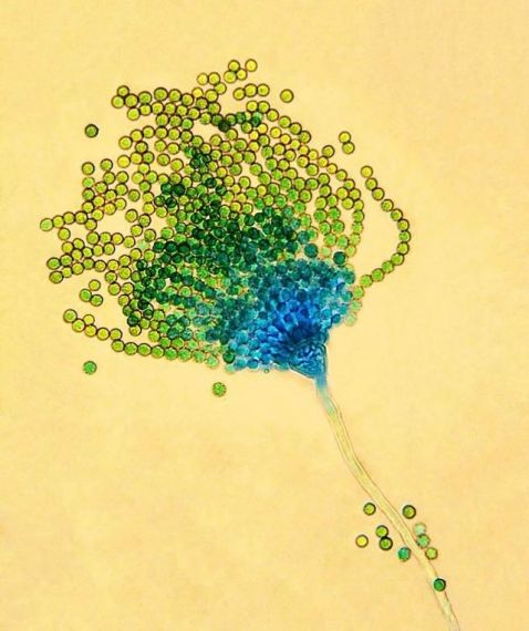
Check out this beautiful Aspergillus parasiticus captured by Tracy Debenport. This fungus was discovered in 1912 after being isolated from mealybugs found on Hawaiian sugarcane plantations. Originally classified as an Aspergillus flavus subspecies due to its similarity, it was renamed due to physiological differences. This sample was stained with lactophenol cotton blue, which Tracy described as giving “peacock feather vibes." We agree! Who knew a parasitic fungus could be so stunning?
Image courtesy of Tracy Debenport. Captured on an Olympus CH-2 microscope.
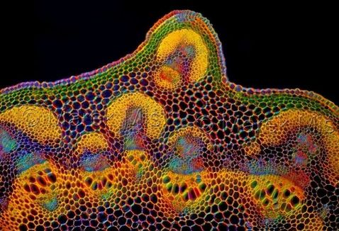
This is a Lathyrus vernus, also known as a spring vetchling or spring pea. This species of flowering herbaceous perennial plant is typically found in Europe and Siberia. It produces bold purple flowers in the spring, which turn greenish-blue as they age.
Image courtesy of Marek Miś. Captured on an Olympus BH2 microscope.
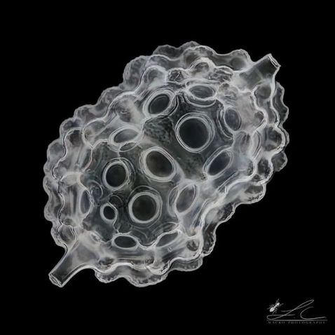
Radiolaria have captivated scientists since the 19th century. Neither animals, plants, nor fungi, these soft-bodied organisms are protists and are notable for their ability to absorb silica from seawater to form elaborate skeletal structures. Found throughout the global ocean and dating back to the Cambrian Period (the first geological period of the Paleozoic Era), the sample captured here is a middle Eocene radiolarian from Barbados.
Image courtesy of Leonardo Capradossi. Captured using inverted colors, brightfield, and image stacking of 172 single shots with an Olympus UPlanSApo objective (40X, 0.95 NA).
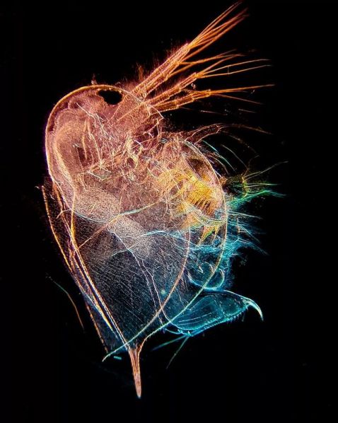
"Daphnia magna is freshwater zooplankton that can grow up to 5,000 micrometers (5 mm). It's the largest species of the genus Daphnia, so it was named D. magna.
Being a keystone species in the freshwater ecosystem, it possesses great tolerance to various conditions. Its transparent abdomen allows scientists to observe its organs nondestructively, making D. magna a great model organism for biological research."
Image and caption courtesy of Minjun Shin. Captured on an Olympus CX21 microscope.
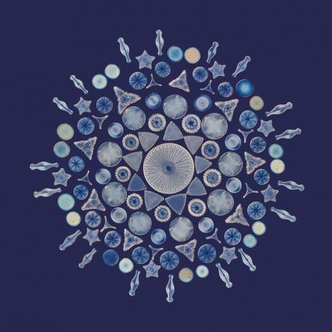
Over a hundred diatoms were arranged into a flower-like circular pattern to make this stunning sample. Microscopists from as far back as the Victorian era were experts in micromanipulation.
According to Smithsonian magazine, diatom “arrangements were often made by professional microscopists… They were sold alongside other miniature curiosities, including microscopic photographs, to wealthy amateur naturalists who would exhibit them at social gatherings as an amusement.”
Image courtesy of Daniel Han. Diatom arrangement by Herbert Potter. Captured by stitching 11 individually stacked images using an Olympus AX70 microscope.
Bonus video—because we all love tardigrades!
This video shows a small tardigrade (also called a water bear) crawling out of its skin. Like snakes, water bears shed their cuticle to protect the eggs they lay within it. As you can see, these three large eggs "bearly" fit in the protective covering.
Video courtesy of Hunter Hines as part of his Tardigrade Takeover. Captured using an Olympus BX53 microscope and filmed with an iPhone 11 Pro under 100X differential interference contrast (DIC).
To see more images like these, be sure to follow us on Instagram at @olympuslifescience.
Interested in sharing your own images? Visit our image submission site.
Related Content
Flying into Summer—Our Most Popular Microscope Images for June 2022
Seeing Shapes—Our Most Popular Microscope Images for May 2022
Awards Season—Our Most Popular Microscope Images for April 2022
