
While you can find beautiful colors somewhere over the rainbow, you can also find them right under your microscope! From brains to bones, this month’s top microscope images boast an array of colors that show a wonderful small world.
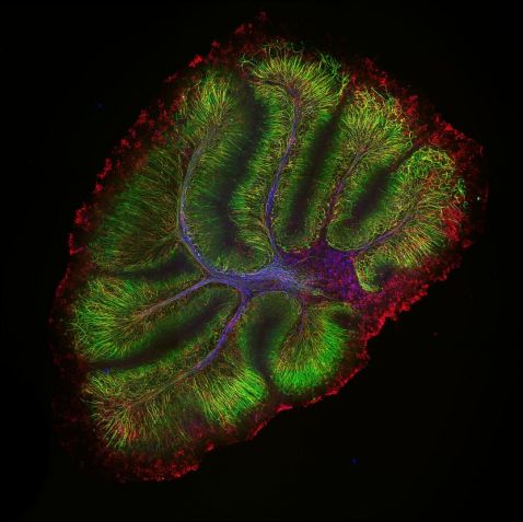
Last month we launched the FLUOVIEW™ FV4000 laser scanning confocal microscope with our breakthrough SilVIR™ detector technology. Captured with the confocal system, this image of a mouse cerebellum is so stunning that it earned a spot in our top five images two months in a row! The cerebellum is known for its coordination and regulation of motor activity, but it also makes for a bright and beautiful image. Neurofilament-heavy chain (NFH) in green, myelin basic protein (MBP) in red, and glutathione S-transferase pi 1 (GSTpi) in blue.
Sample courtesy of Dr. Katherine Given, Principal Investigator, Neurobiology, University of Colorado Anschutz Medical Campus, Aurora, Colorado. Image captured on a FLUOVIEW FV4000 laser scanning confocal microscope using an UPLXAPO10X X Line™ objective.
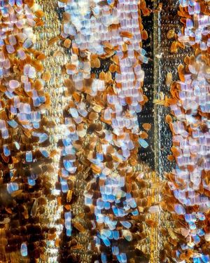
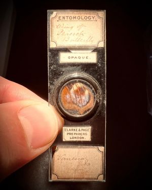
This focus-stacked image is of a butterfly wing from an Aglais io, also known as the peacock butterfly. Found in Europe and temperate Asia as far east as Japan, this specimen is from a slide made about 100 years ago. Both time and butterflies fly, but this sample is still as fascinating today as it was when it was originally created!
Image courtesy of Brian, @brinybrine. Captured on an Olympus BH-2 microscope.
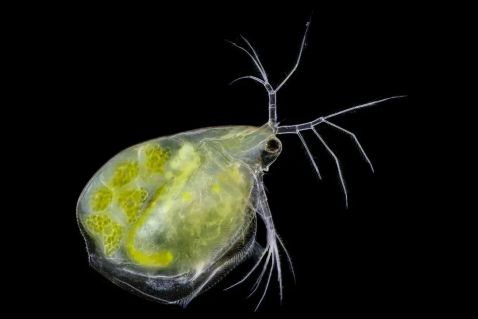
It's amazing how even the tiniest of creatures evolve and adapt to their environments, like this Daphnia.
"Last year, I noticed something strange: Daphnia had some parasitic fungus inside. Today, there aren't any parasites, but the species had changed. Natural selection at its best! Here in Italy, the storm Cirian did lot of damages to beach structure, which is why you don't see any plankton. That is also natural selection, as you cannot build on the beach and expect nothing would happen. However, enjoy one of the best photos I took during the storm yesterday!"
Image and caption courtesy of Luigi Bozzano. Captured using an Olympus 10X UPlanApo objective.
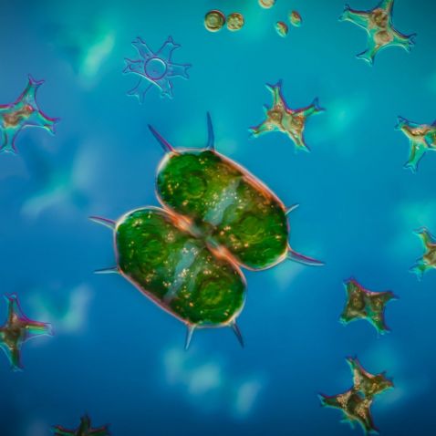
We’re in love with this Xanthidium antilopaeum (posing in front of some friends of unknown species). Xanthidium, a green alga, exists as symmetrical single cells, which form two halves called semicells. This sample was captured from the freshwater ponds at Drottningholm in Stockholm, Sweden.
Image courtesy of Håkan Kvarnström. Captured on an Olympus BX51 microscope with differential interference contrast (DIC) and using a SAPO60X water immersion objective.
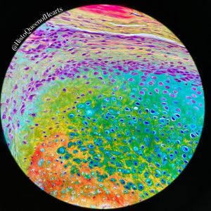
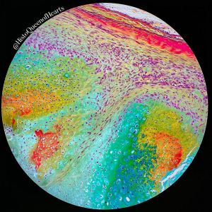
Katelin Murphy may be known as the queen of hearts, but we think she’s the queen of colors! These images captured of a pentachrome stain are bursting with a rainbow of hues.
“This method is used for the demonstration of collagen, elastin, muscle, mucin, and fibrin in tissue sections. The vast array of colors make for some very beautiful stains, especially in this section of bone.”
Image and caption courtesy of Katelin Murphy. Captured on an Olympus BH40 microscope.
Bonus video! These ginkgo leaves put the Y in ROYGBIV, the acronym for the seven colors of the rainbow: red, orange, yellow, green, blue, indigo, and violet.
"The vein pattern of the ginkgo leaf is so unique and beautiful. No other plant has such a vein pattern. This is because ginkgos are living fossils. They are called living fossils because they haven’t changed much in the millions of years since they first appeared on our planet.
Looking at the ginkgo leaf in more detail, you can now see the arrangement of the cells that make up the leaf veins as well as the pores (stomata) that the leaves use to take up CO2 and release O2.
But the most amazing thing about the leaves that I was looking at? They are full of fungi growing on them! These fungi are difficult to see under normal conditions because they are colorless and the color of the leaf is intense. But when I used the dye methylene blue, the fungal hyphae was stained, and now you can clearly see the fungi."
Video and caption courtesy of Adolfo Sánchez-Blanco, PhD. Captured on an Olympus CX31 microscope.
To see more images like these, be sure to follow us on Instagram at @evidentlifescience!
Interested in sharing your own images? Visit our image submission site!
Related Content
Pretty Pollen—Our Most Popular Microscope Images for October 2023
Magnificent Microorganisms—Our Most Popular Microscope Images for September 2023
Breadth of Beauty—Our Most Popular Microscope Images for August 2023
