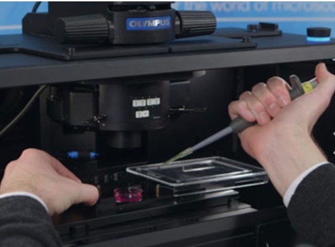
Do you have new long-term live cell or model organism research applications but aren’t sure if your older inverted research microscope can handle them? Is the cost of a new inverted microscope outside your current budget? You may be surprised to learn that with a few key modifications your current microscope may still have “the legs” to achieve your research aims.
Along with the upgrades discussed in Part 1 and Part 2 of our microscope improvement series, adding an environmental control chamber or enclosure could help you achieve live imaging research goals you may not have considered feasible.
The Importance of Environmental Control for Stable Live Cell Imaging
Live cell imaging may take 20 minutes, several hours, or even longer. In addition to Z-focus drift associated with room temperature changes in longer experiments, you may need to control for:
- Temperature (up to 37 °C [99 °F] or down to 22 °C [72 °F])
- Humidity
- CO2 (and O2 if hypoxic conditions are relevant)
Microscope environmental control can be configured to include any or all of these capabilities, making it an easy upgrade for various live cell and model organism imaging applications.
The Benefits of Stage-Top Units
Stage-top systems such as those from Tokai Hit and Okolab offer conveniently removable chamber units. This enables the addition and removal of environmental control as needed. This is particularly convenient when the instrument is also used for applications that don’t require live cell imaging.
Moreover, these systems are highly effective at maintaining viability for hours and even days. Perfusion ports enable water and media replenishment for humidity and nutrients/agonists, and available in-line solution heaters minimize thermal shock.
Please also note that immersion objectives act as heat-sinks when in contact with your sample, so you may want to add an objective heater if using these optics.
Microscope Enclosures for Live Cell Imaging
Another option is a full microscope incubation enclosure. While enclosures can’t be easily removed from the microscope, they can maintain environmental control within a larger volume than with a stage-top chamber.
These enclosures are spacious enough to accommodate fluid dispensation rigs and provide stable temperature environments. This is beneficial for both the sample and the microscope as frequent hot/cool cycles can increase wear and tear on microscope components. Many manufacturers offer a blackout model to maintain a dark environment within the enclosure, eliminating the need for a separate microscope room.
Impact of Changes in a Room Environment on Time-Lapse Imaging
It’s important to note that most microscope incubation systems are calibrated for an average temperature of 25 °C (77 °F). Some systems precisely monitor temperature and make rapid temperature corrections. Even so, it’s best to avoid placing your instrument in areas subject to thermal changes. Remember to avoid placing the instrument under vents, but if this is unavoidable, use baffles to direct airflow away from the microscope.
Finally, make sure the microscope is located away from large instruments such as centrifuges, refrigerators, freezers, and frequently used doors. These can impart vibration artifact into your time-series imaging experiment. A pneumatic antivibration table is helpful for isolating your instrument, but it’s best to avoid risking any vibrational disruption in your imaging experiment.
Get More Out of Your Microscope
Want more microscope improvement tips? Don’t forget to review Part 1 and Part 2 of this series to discover other system updates to consider for effective live cell imaging. Check them out now:
- 5 Ways to Extend the Life (and Performance) of Your Microscope
- How to Make Your Microscope Light Source Last Longer and Perform Better
Related Content
5 Ways to Extend the Life (and Performance) of Your Microscope
4 Tips to Achieve Longer Live Cell Imaging with Less Time in the Lab
How to Make Your Microscope Light Source Last Longer and Perform Better

