Hoffman modulation contrast, also known as relief contrast, is an optical microscopy technique designed to increase the contrast of unstained and living material contained in a plastic vessel. It works by detecting optical gradients and converting them into variations of light intensity.
This post explores Hoffman modulation contrast in detail—from its basic microscope configuration to its advantages and limitations.
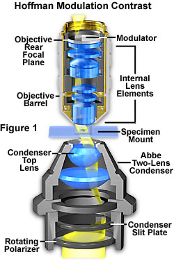
Figure 1 Basic microscope configuration for Hoffman modulation contrast
Basic Microscope Configuration for Hoffman Modulation Contrast
Figure 1 shows the basic microscope configuration for Hoffman modulation contrast. An optical amplitude spatial filter, termed a "modulator" by Hoffman, is inserted on the back focal plane of an objective. Light intensity passing through this system varies above and below an average value, which is then modulated. Objectives useful for modulation contrast can cover the entire magnification range of 10X, 20X, and 40X.
The modulator has three zones (depicted in Figure 2):
- A small, dark zone near the periphery of the back focal plane transmits only one percent of light (areas marked D in Figure 2)
- A narrow gray zone transmits 15 percent of light (areas marked G in Figure 2)
- The remaining clear or transparent zone, covering most of the area at the back of the objective, transmits 100 percent of the light (areas marked B in Figure 2)
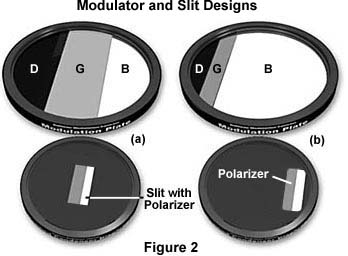
Modulator and slits for Hoffman modulation contrast microscopy
Unlike the phase plate in phase contrast microscopy, the Hoffman modulator is designed not to alter the phase of light passing through any of the zones. When viewed under modulation contrast optics, transparent objects that are essentially invisible in ordinary brightfield microscopy take on a three-dimensional (3D) appearance dictated by phase gradients.
In the Hoffman design the slits are at the front focal plane of the condenser, as illustrated in Figure 1. When light passes through the off-axis slit, it is imaged at the back focal plane of the objective (also termed the Fourier plane) where the modulator is installed. The front focal plane of the condenser containing the off-axis slit plate is optically conjugate to the modulator in the objective back focal plane. Image intensity is proportional to the first derivative of the optical density in the specimen and is controlled by the zeroth order of the phase gradient diffraction pattern.
Opposite gradients result in deflection of the slit image to either the very dark part or the bright section of the modulator. Consider this example: a hypothetical specimen containing both positive and negative phase (thickness) gradients and a flat (non-gradient) area is imaged using modulation contrast optical components. The positive gradient deflects light into the clear area of the modulator, where it is not attenuated, and 100 percent of this light transmits into the intermediate image plane. Likewise, light deflected by a negative gradient into the dark area of the modulator is attenuated to approximately one percent of its former value.
Any non-gradient part of the specimen and the background register on the gray part of the modulator, where about 15 percent of the light transmits into the intermediate image plane. The result is that the intensity of the image area from one side of the gradient is dark. Intensity from the opposite side of the gradient yields a bright image area, and the non-gradient areas appear gray on the image, as does the background.
4 Key Advantages of Hoffman Modulation Contrast
There are numerous advantages and some limitations to Hoffman modulation contrast. Some advantages of Hoffman modulation contrast include:
1. Fuller use of your objective’s numerical aperture.
The ability to use higher numerical apertures with Hoffman modulation contrast yields excellent resolution of details along with good specimen contrast and visibility.
2. Works with plastic vessels.
Hoffman modulation contrast can be successfully used with birefringent materials, such as plastic dishes, without image distortion. Birefringence, or double refraction, is the phenomenon exhibited in certain materials where an incident ray (a ray of light that strikes a surface) is split into two rays when it passes through the material.
Hoffman modulation contrast a good alternative to contrast methods that do not work well with birefringent materials. For instance, examining birefringent materials with differential interference contrast (DIC) can lead to artifacts, loss of contrast, and other image quality issues.
As a result, Hoffman modulation contrast is an ideal method for observing and imaging cell, tissue, and organ culture in plastic containers.
3. The ability to perform optical sectioning.
It is also possible to do "optical sectioning" with this technique. Sectioning enables you to focus on a single thin plane of the specimen without interference from confusing images arising in areas above or below the plane that is being focused on.
The depth of a specimen is measured in a direction parallel to the optical axis of the microscope. Focusing the image establishes the correct specimen-to-image distance, allowing interference of the diffracted waves to occur at a pre-determined plane (the image plane) positioned at a fixed distance from the eyepiece. This enables diffracting objects that occur at different depth levels in the specimen to be viewed separately—provided there is enough contrast.
The entire depth of a specimen can be optically sectioned by sequentially focusing on each succeeding plane. In this system, depth of field is the distance from one level to the next where imaging of distinct detail occurs and is controlled by the objective’s numerical aperture. Higher numerical aperture objectives exhibit very shallow depths of field. The opposite holds for objectives of lower numerical aperture. The overall capability of an objective to isolate and focus on a specific optical section diminishes as the optical homogeneity of the specimen decreases.
4. Enhanced visibility.
Another advantage of Hoffman modulation contrast is enhanced visibility. Images appear shadowed or pseudo three-dimensional, enhancing visibility because of differences in contrast on either side of a detail. There are also no halos exhibited in the images—unlike in images produced with phase contrast optics. Hoffman modulation contrast converts phase gradient information into amplitude differences that are very different from the phase relationship variations (and optical path differences) produced by a phase contrast microscope.
The use of the dark and gray zones in the modulator produces images that contain varying shades of gray and are devoid of color. It is possible to introduce color into modulation contrast images by producing modulators with the gray and dark zones substituted for colored zones of equal transmittance values. In this case, the resulting images from the phase gradient would render in colors with similar gradients in the same tint. Currently, we are unaware of any commercial source of modulation filters that contain colored zones.
Limitations of Hoffman Modulation Contrast
There are also several disadvantages and limitations of Hoffman modulation contrast. First, these images must be viewed with caution. The reason is that different observers can see a "hill" in the image as a "valley" or vice versa since the pseudo three-dimensional image is observed through the eyepiece. The system is also highly sensitive to gradients perpendicular to the length of the slit. As a result, this technique requires some degree of skill to position the specimen for the best effect.
Related Content
Stereo Microscope Primer: Transmitted Light Observation Methods
Microscopy Solutions for Assisted Reproductive Technology (ART) Research