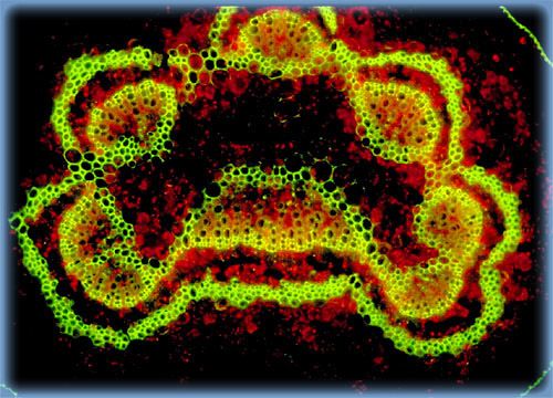
Japanese Maple Leaf Petiole
A fluorophores-stained cross section of a leaf petiole or leaf stalk from a Japanese maple (Acer palmatum) was examined utilizing widefield fluorescence microscopy to capture both secondary and autofluorescence emitted by the leaf tissues. The photomicrograph presented below reveals conducting tissues that transport nutrients and other materials to and from the leaf.
Sorry, this page is not
available in your country.