2022年EVIDENT全球显微图像大赛我们激动地宣布2022年Evident全球显微图像大赛的获奖者名单。我们感谢每一位参赛者,并期待您明年再次参加。 |
|
全球奖得主这张荣获全球奖的图像由Laurent Formery(美国)拍摄。 图像显示的是宽约1厘米的幼年海星(Patiria miniata)的神经系统。样品在经过透明化处理后用抗乙酰化微管蛋白的抗体进行标记,并使用颜色编码的Z轴投影展示。 | 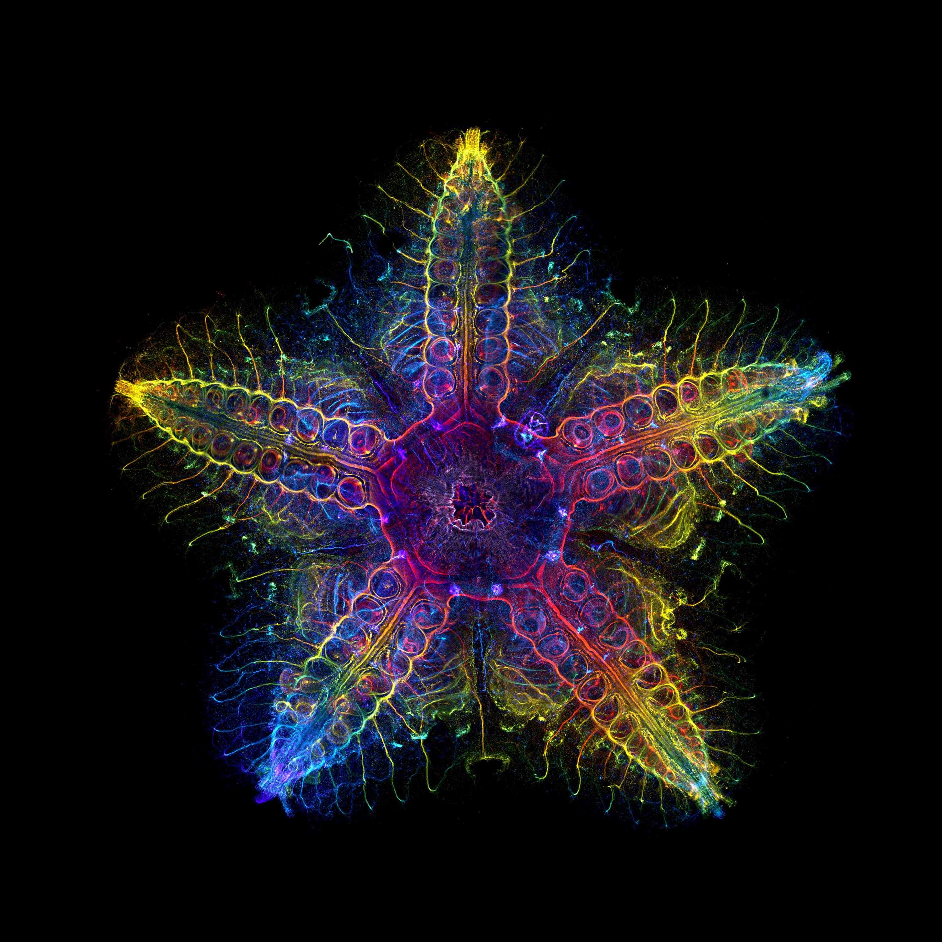 |
|---|
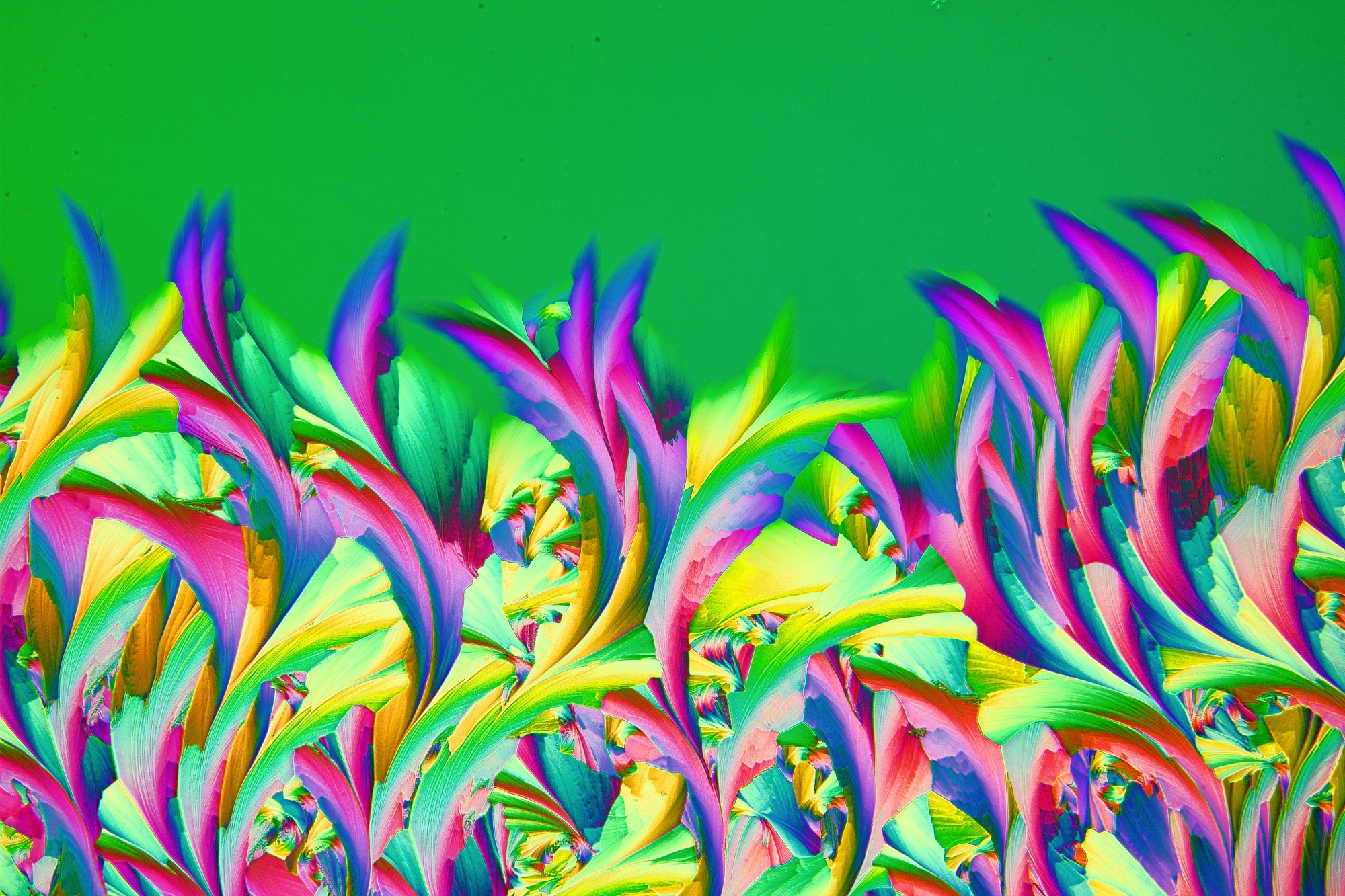 | 材料科学奖得主材料科学类获奖图像由Shyam Rathod(印度)拍摄。 图像显示的是一种名为ABE的治疗疣的外用药物结晶(这种药在波兰有售)。露水被吸管吹到显微镜载玻片上,并被拍摄成单幅图像。使用了延迟器和双交叉偏振滤光片来突出色彩。 |
|---|
区域奖得主
美洲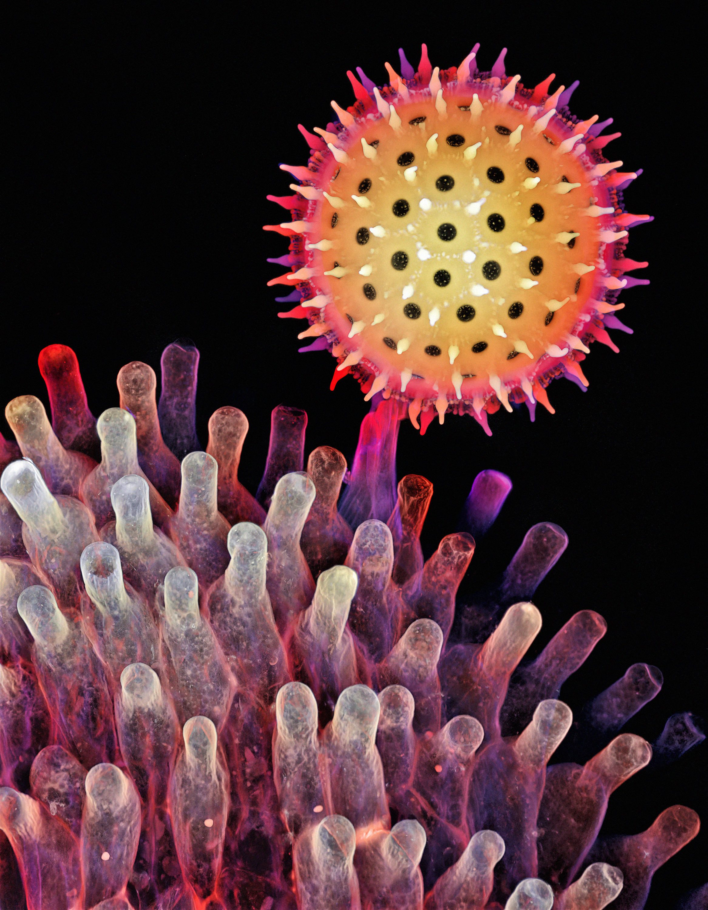 美洲地区的获奖图像由Igor Siwanowicz(美国)拍摄。 以深度彩色编码投影展示的牵牛花附着在柱头上的发芽花粉粒。 | 欧洲、中东和非洲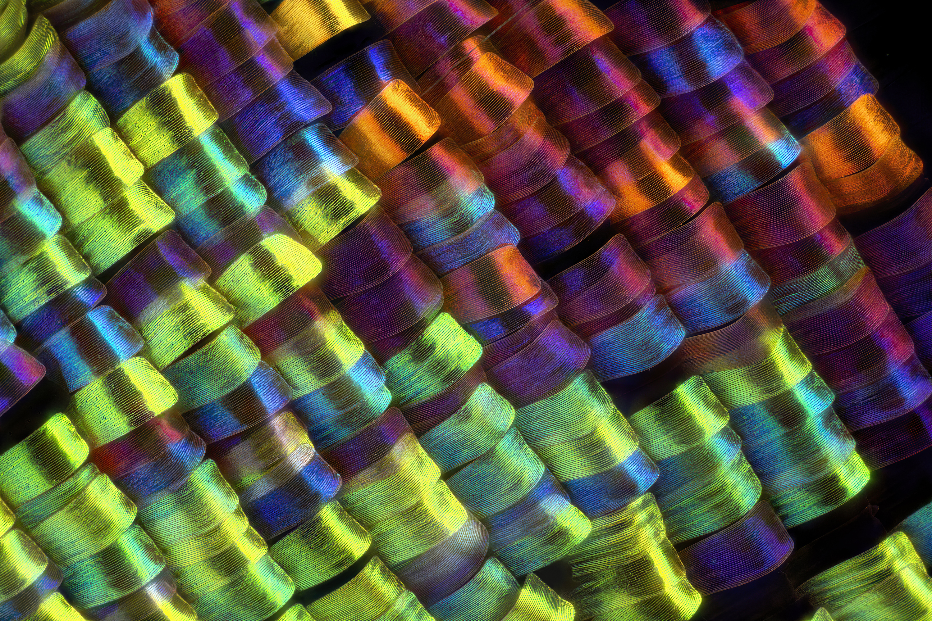 欧洲、中东和非洲地区(EMEA)的获奖图像由Javier Ruperez(西班牙)拍摄。 图像显示的是Urania rhipheus蝴蝶翅膀上的鳞片。在20X倍率下拍摄。 | 亚太地区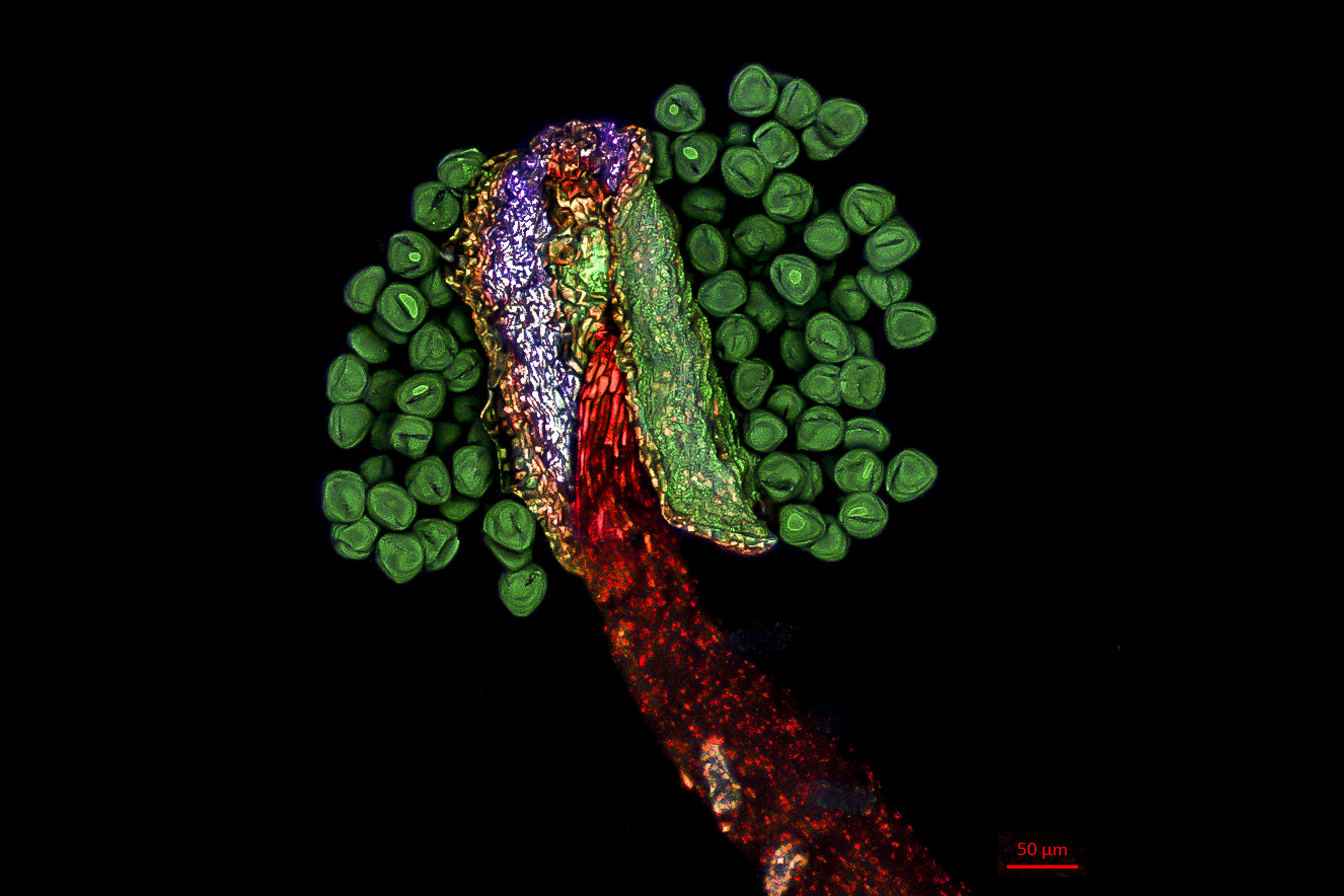 亚太地区的获奖图像由Jiao Li(中国)拍摄。 图像显示的是雪绒花雄蕊,由激光扫描共聚焦显微镜进行三维扫描和重建。 |
荣誉奖
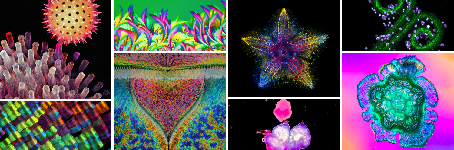
 受精后72小时的斑马鱼的外侧头。 |  红背蝾螈头骨的背视图。采用莫瓦特五色染色技术染色。 |  硅藻是一种单细胞生物,可在世界各地的海洋、水沟和土壤中找到。 |  成年大鼠离体心肌细胞的三色共聚焦成像,分别显示细胞核、溶酶体和线粒体。 | 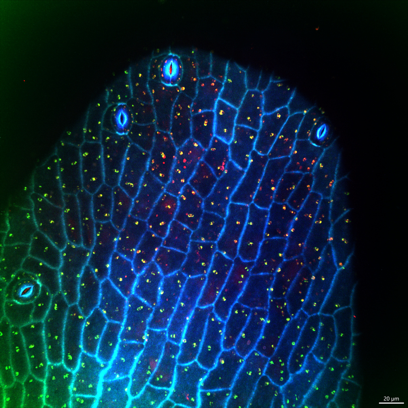 花朵雄蕊顶端在405 nm、488 nm和561 nm波长下的自发荧光,发出孔雀羽毛般的青蓝色和翠绿色。 |
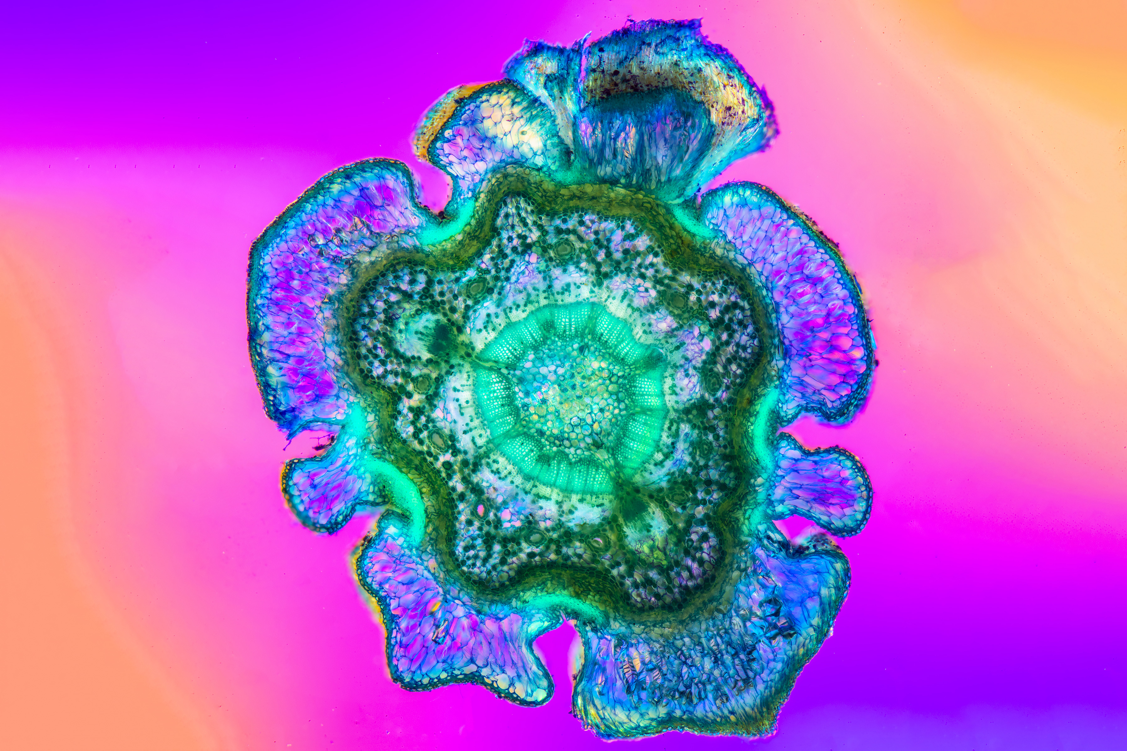 蓝云杉(Picea pungens)枝条的横截面。此图像是使用5倍物镜拍摄并对多张照片进行聚焦扩展处理而得到的全景图。 | 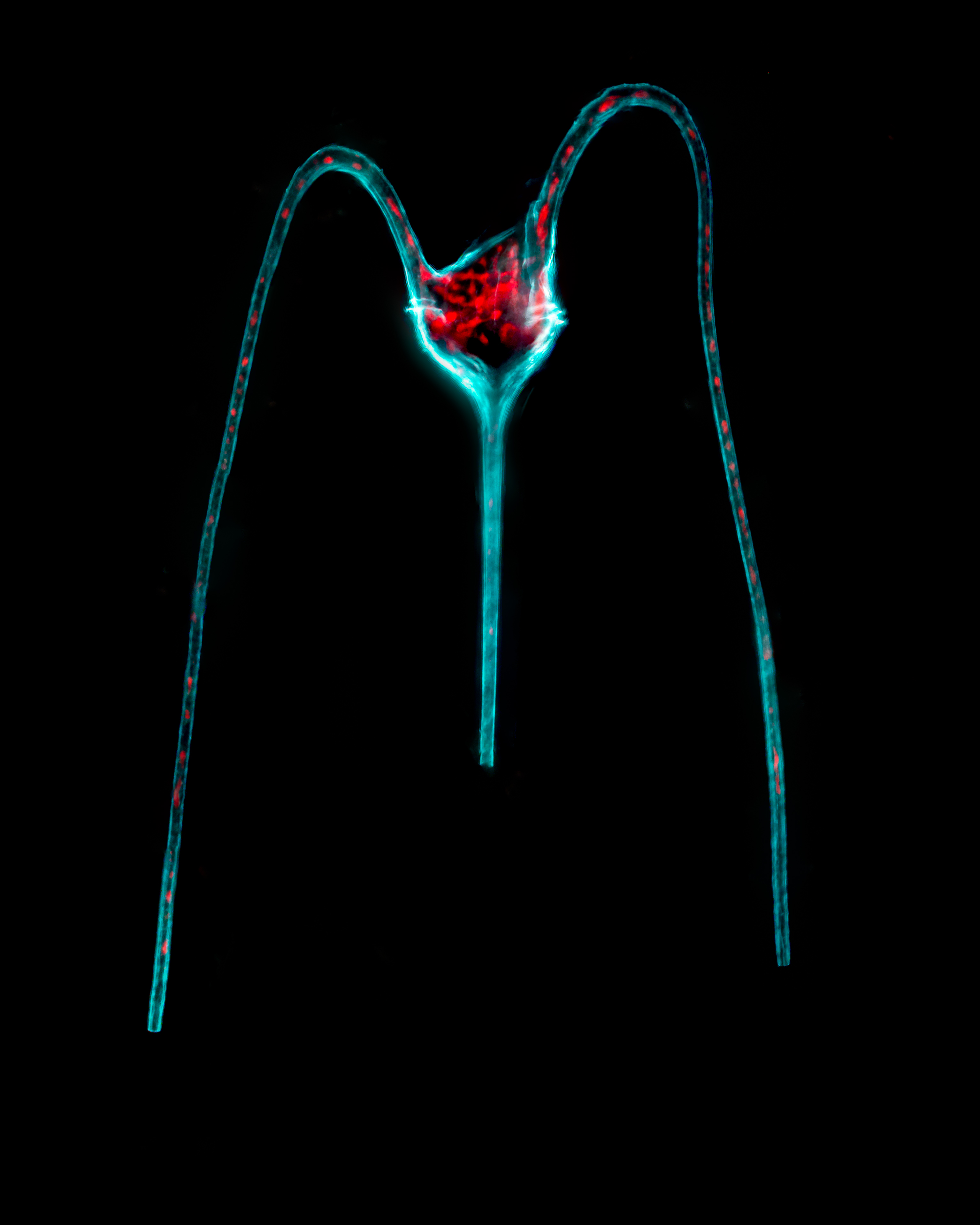 三角大角藻。一种长有三个角的单细胞微藻。其上面的两个角弯向下面的单顶角。叶绿素显示为红色,这是叶绿体所在的位置。 | 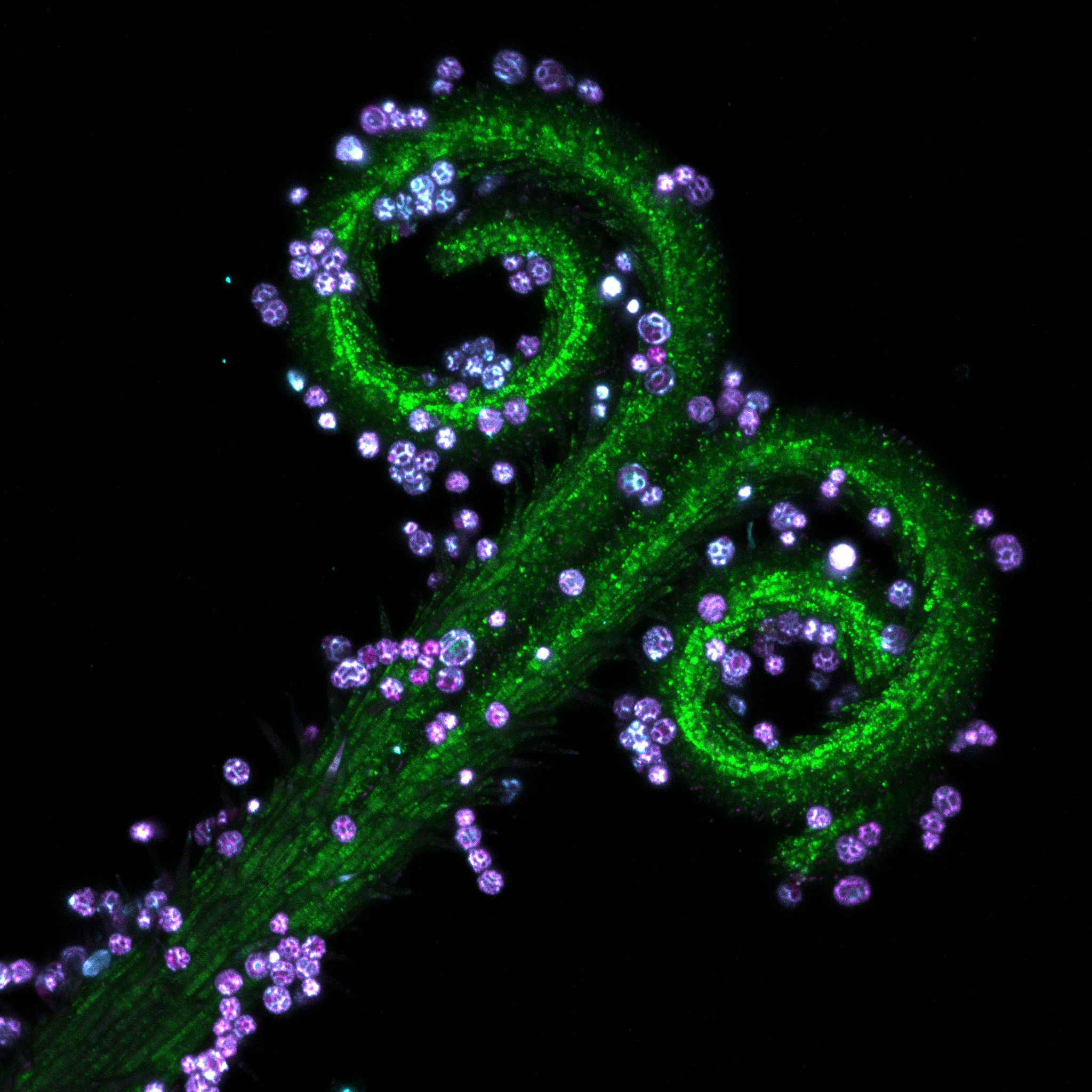 将采集的蒲公英雄蕊制成玻片样品。使用共聚焦显微镜观察其各部分的不同荧光图案。 | 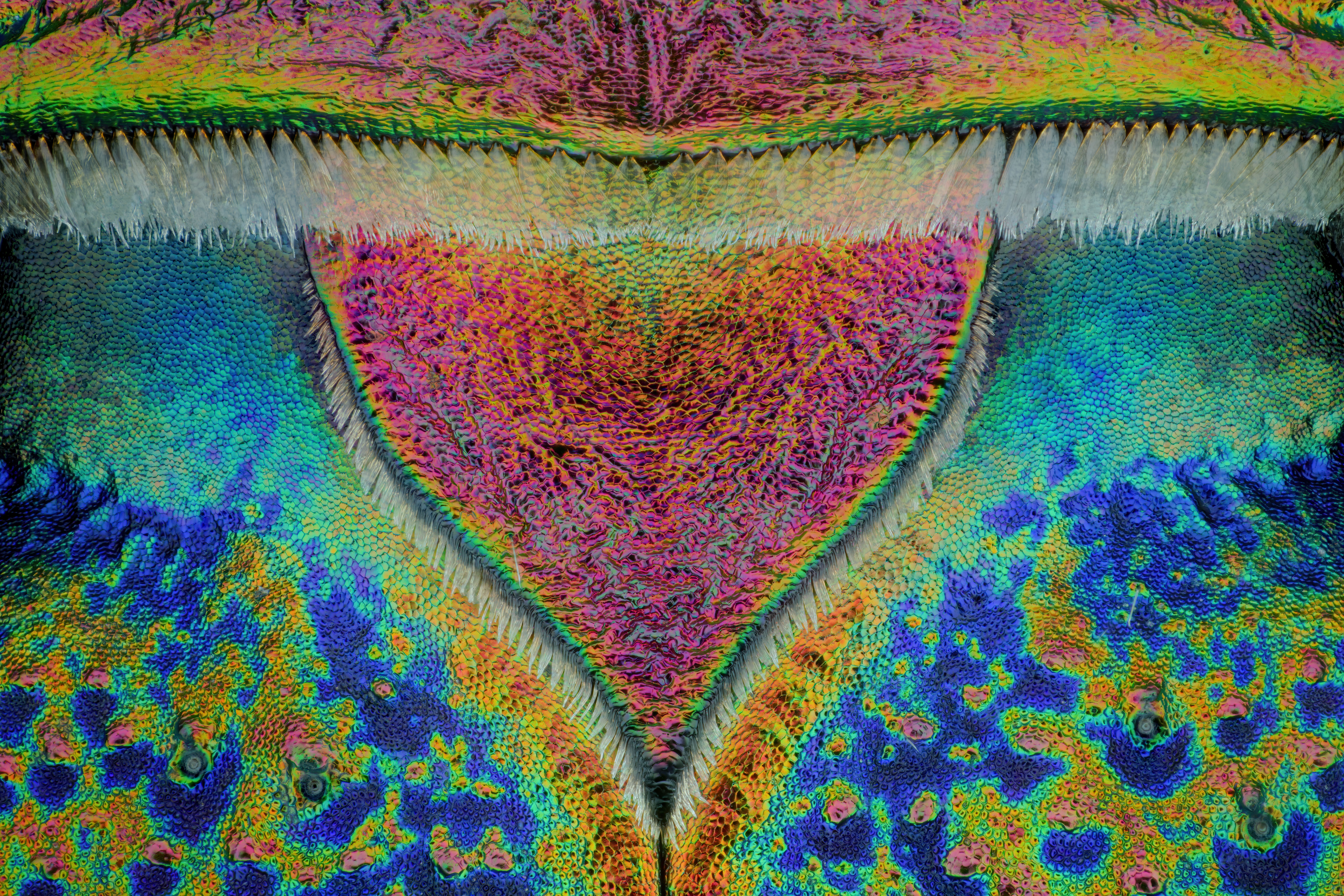 虎甲虫的盾片20:1 | 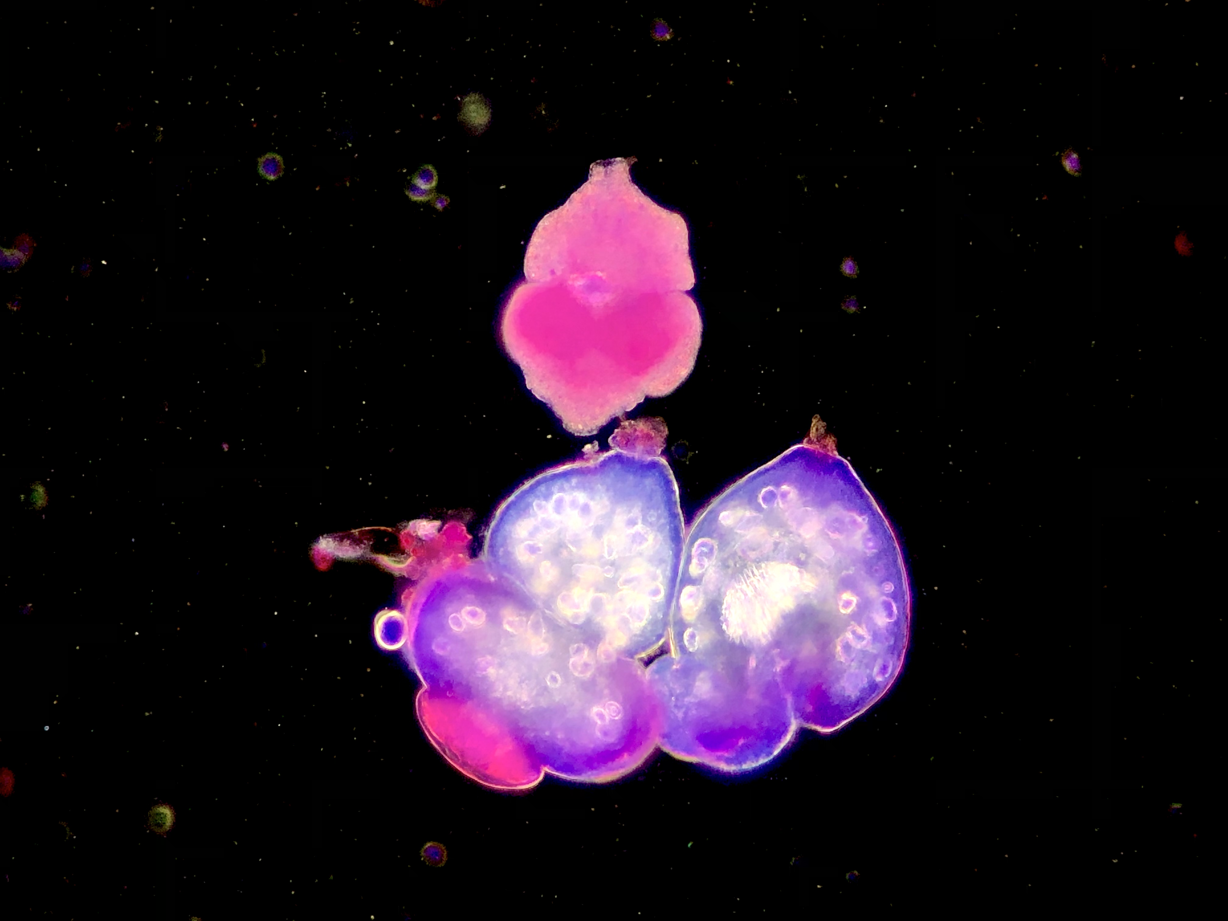 细粒度原头棘球绦虫,用SM染色,并进行暗场拍照。 |
评委
 | Geoff Williams,美国布朗大学Leduc生物影像平台负责人Geoff Williams在布朗大学Leduc Bioimaging Facility(生物影像平台)任职经理已达十四年。早在康涅狄格学院攻读本科学位期间,他已经借助电子显微镜和光学显微镜的发现,将视觉艺术、科学、技术与精湛的技能结合在一起。Geoff从密歇根州立大学研究生毕业后,曾参与运营中央密歇根大学影像平台,并最终担任布朗大学影像平台经理。在过去的20多年中,他一直在磨练自己的电子和光学显微镜技艺。比起纯粹的科学探索,他更注重每幅图像的美学。Geoff以“Nanoscape”命名的作品集收藏了他多年以来积累的形态各异、令人震撼的显微图像,为我们展示了在日常生活中不太可能见到的微观世界。 |
 | Harini Sreenivasappa,德雷塞尔大学细胞成像中心的负责人Harini Sreenivasappa是德雷塞尔大学细胞成像中心光学显微镜核心平台的经理。在德州农工大学(TAMU)研究生院就读期间,她接触到显微镜,当时她研究的课题是微环境刺激在心血管疾病血管壁重塑中对细胞感知和适应的作用。她由此获得了生物医学工程的博士学位。她在使用各种显微镜技术方面拥有超过10年的丰富经验,例如原子力显微镜(AFM)、转盘式共聚焦显微镜和全内反射荧光(TIRF)显微镜。借助美国细胞生物学学会(ASCB)的COMPASS Outreach资金,她创立策划了“旅行显微图像”展览,展出了德州农工大学(TAMU)研究人员拍摄的显微图像,并免费向公众开放。该系列展览的目的是与当地社区分享德州农工大学的研究成果,并激发人们对成像科学的兴趣。 |
 | Rachid Rezgui,纽约大学阿布扎比分校,显微镜研究仪器科学家Rachid Rezgui是一位显微镜专家,也是一位活跃的研究科学家。Rachid曾在德国汉诺威莱布尼兹大学学习物理学,随后在法国综合理工学院研究单分子水平上的DNA-蛋白质的相互作用,并获得生物物理学博士学位。2014年,他加入纽约大学阿布扎比分校的显微镜成像中心,此后,他在工作中使用过所有类型的显微镜,包括双光子、超分辨率、共聚焦、荧光寿命、宽场等。他的工作涉及到光学成像的各个方面,如样品制备、培训、采集、后处理以及成像中心的管理。 |
 | Urs Ziegler,苏黎世显微镜和图像分析中心常务主任Urs Ziegler于1996年在苏黎世大学生物化学研究所获得生物化学博士学位。2007年,他成为显微镜和图像分析中心的负责人。他的研究包括开发和引进各种用于多细胞生物结构研究的低温电子显微镜方法、超分辨率光学显微镜以及新的光电联合的显微成像方法。研究之余,他还教授高级显微镜和图像分析方面的各种讲座和实践课程。自1998年以来,他一直是瑞士光学和显微镜学会的理事会成员。 |
 | 孙育杰,北京大学终身教授、博雅特聘教授孙育杰博士在中国科技大学获得化学学士和硕士学位,在匹兹堡大学获得化学博士学位。随后,他作为博士后研究员加入宾夕法尼亚大学医学院,与一个跨学科团队合作,解决分子马达如何使用单分子荧光工作以及操纵技术的难题。孙育杰博士是未来技术学院(CFT)的副院长,纳米/生物界面中心(NBIC)副主任,以及国家多模态跨尺度生物医学成像中心的副总工程师。他任职于许多专业科学组织,包括美国细胞生物学会(ASCB)、生物物理学会(BPS)和中国生物物理学会(BSC)。孙育杰博士一直在开发先进的单分子成像和操纵技术来研究细胞结构和功能。他的研究成果发表在5部专著及100多篇同行评议论文中,他先后承担了10个国家级科研项目。 |
 | Sarah Ellis,Olivia Newton-John癌症研究所肿瘤环境成像中心(CITE)副教授Sarah Ellis拥有30多年从事各种显微镜工作的丰富经验,在光学和电子显微镜的样品制备、图像数据分析以及宽场、共聚焦、多光子和生物电子显微镜的操作和维护方面拥有精湛的技能。Sarah在核心设施设计和管理方面也拥有丰富的经验。她喜欢培训研究人员,并拥有强大的协作网络,从她作为作者参与了60多篇出版物的事实就可证明这点。Sarah通过从事多项志愿工作为科学界做出了贡献,她是澳大利亚显微镜和显微分析协会的秘书,也是澳大利亚光学显微镜协会的维多利亚州代表。 |
下载壁纸
免费下载2022年显微图像大赛壁纸包来美化您的屏幕吧!
虚拟背景下载可在虚拟会议中使用的背景。 | 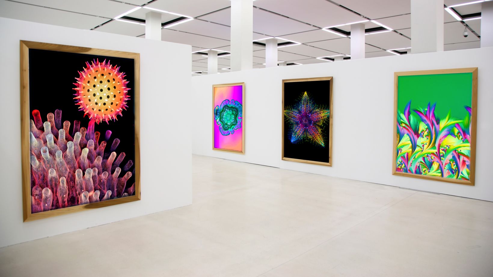 |
往届获奖者
|
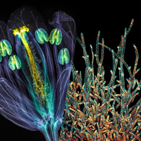
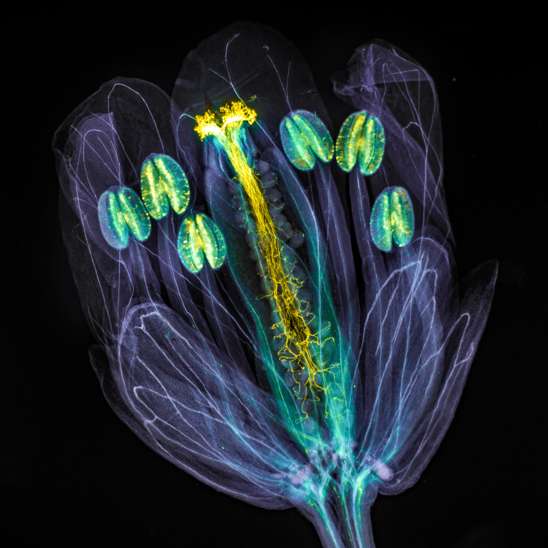
荣获全球奖的图像由Jan Martinek(捷克共和国)拍摄。 拟南芥花,花粉管从雌蕊中生长而出。花朵组织经过化学处理后变得透明,并使用苯胺蓝(黄色荧光)对花粉管进行染色,以便于观察。 |
对不起,此内容在您的国家不适用。
对不起,此内容在您的国家不适用。