
Love is in the air and shows up in our samples. Check out our roundup of popular microscope images (and microscopic valentines) for February.
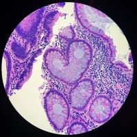
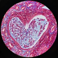
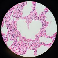
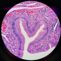
We love the concept of pareidolia: seeing familiar objects or shapes in unrelated objects or patterns. Here we see a series of H&E stains with heart shapes fit for a Valentine’s Day card. These beautiful samples include a human colon, dog uterus, goat lung, and lizard gastrointestinal (GI) tract.
Images courtesy of Kate Murphy. Captured using an Olympus BX40 microscope.
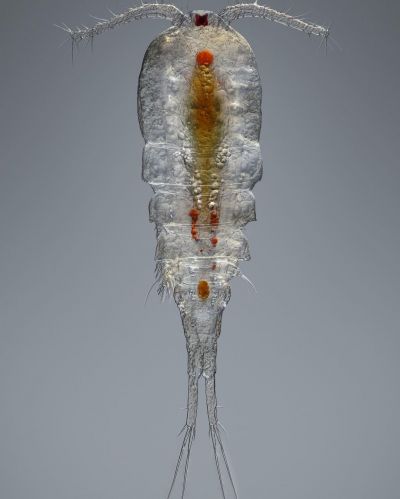
Did you know that copepods are fast swimmers? Some copepods can travel distances of 295 feet (90 m) in an hour. This is the human equivalent of swimming 50 miles per hour (81 km/h).
Here is a full-body look at a female cyclopoid copepod found in a pond in the Dubai desert.
Image courtesy of Leonardo Capradossi. Focus-stacked image captured at 100x magnification using an Olympus 10X UPLANFLN objective lens, 0.30 NA; oblique illumination.
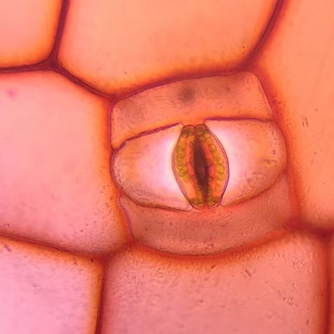
This next image is another great example of pareidolia. This stoma in a plant cell appears to be an eye looking at us as we look at it!
Image courtesy of Eva Petrovova. 2019 Image of the Year Award submission. Captured using an Olympus CX23 microscope.
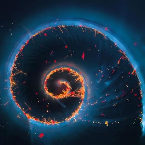
It's no wonder this stunning image won first prize in our 2018 Image of the Year contest and consistently reappears as a favorite! Håkan Kvarnström perfectly captured the shell of a marine snail covered in algae and cyanobacteria. This small, 2 mm snail was found at the bottom of a bottle of water collected from a local fish farm in Stockholm, Sweden. To learn more about the image, check out our spotlight on Håkan.
Image courtesy of Håkan Kvarnström. 2018 European Image of the Year Award winner. Captured using an Olympus BX51 fluorescence microscope.
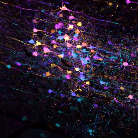
This image shows a tissue section of a mouse brain that was labeled with trans-synaptic viral tracer to investigate neural pathways that link the visual systems in the mouse. The varying colors are beautiful and serve a purpose by reflecting the changing depths of neuronal processes.
Image courtesy of Arthur Chien. Captured using an Olympus FV3000 confocal microscope.
Bonus video!
This determined nematode (roundworm) is trying its best to enter an air bubble located on the slide. Nematodes outnumber all other animals on Earth, making up roughly 80% of all animals we can find. These small, abundant worms play an important role in our environment by accelerating the decomposition process. Their feeding recycles minerals and other nutrients from bacteria, fungi, and plants back into the soil. Nematodes are also sensitive and responsive to changes in the environment caused by pollution.
Video and caption courtesy of Håkan Kvarnström. Captured on an Olympus BX51 microscope.
To see more images like these, be sure to follow us on Instagram at @olympuslifescience.
Interested in sharing your own images? Visit our image submission site.
Related Content
Diatoms to Dinosaurs—Our Most Popular Microscope Images for January 2022
Aesthetics of Autofluorescence—Meet the IOTY 2020 Global Winner
Surf and Turf—Our Most Popular Microscope Images for December 2021
