The period to enter your microscopy images for the Evident 2022 Image of the Year Award is winding down with just a few weeks to go. You can find the deadline and the contest details on the IOTY 2022 submission page.
Every year, we profile the winners—one global winner and 3 regional winners—but this year we thought it’s high time we put our amazing jurors under the microscope. Our IOTY competition depends on the participation of our esteemed and knowledgeable jurors from around the globe. We are grateful for their willingness and enthusiasm, and a little in awe. What motivates them to contribute their time and expertise to judging our competition?
We interviewed these 5 jurors, past and present, to find out why they’re so passionate about microscopy imaging. We also delve into what they think constitutes a winning micrograph. Read on to get some valuable tips.
Meet 5 EVIDENT IOTY Award Jurors


Q: Where and when did you first learn to use a microscope?
Stefan: As a teenager I was already interested in light microscopy and my wish to have my own light microscope was fulfilled as a Christmas present. My learning was restricted to self-education at that time.
Sian: I first learned to use a microscopy as part of a Physiological Society summer studentship during my undergraduate studies at UCL. I was working in Jonathan Ashmore’s lab, investigating calcium signaling in inner hair cells in the cochlea.
Geoff: Growing up, I was given a small Edmund Scientific plastic compound microscope kit, and I looked at a lot of pond water. It wasn’t until college when I first started with more complicated microscopes. The first research level system was an Olympus upright fluorescence system with a film camera. That system and the old SEM/TEM combo were the first exposure to the next level of imaging, combining it with my art foundation seemed natural. From there, grad school and beyond, and it has been microscopes ever since.
Rachid: During my master’s thesis at the Laser Zentrum Hannover in Germany.
Harini: I was first introduced to a microscope during high school biology class where we used a compound microscope to observe the cross section of a plant stem and root. I first learned how to use a microscope while pursuing the biomedical engineering PhD program at Texas A University under the guidance of Dr. Andreea Trache.
Q: When did you begin using microscopic imaging as part of your career?
Stefan: My choice to focus on microscopy was mainly triggered by the practical microscopy methods course organized by Professor Schnepf and colleagues, which I took as a biology student during my studies at University of Heidelberg.
Harini: I have always been passionate about applying engineering methods to better understand the mechanisms of disease progression. My interest pivoted toward a career in microscopy during my later years of graduate school. The combination of interdisciplinary training in cell biology, electrical and biomedical engineering, along with proficiency in microscopy and a passion for teaching organically led me to a career in microscopy core facility management.
Sian: I’ve been using microscopy throughout my whole career in various forms. In my first microscopy project, I was very much focused on using microscopy to study a biological process, whereas in my PhD I was looking more at developing new microscopy techniques from an optical physics and spectroscopy perspective. In my postdoc, and now my own group, I am interested in using computational analysis to process and extract information from microscopy images. Basically, if something involves microscopy, I’m interested!
Geoff: When I wanted to do plant development research, microscopy was a very natural part of the career path. But graduate school opened me up to the realization I could look at everything and make microscopes the focus, both in understanding how the work, fixing them, and teaching people how to use them.
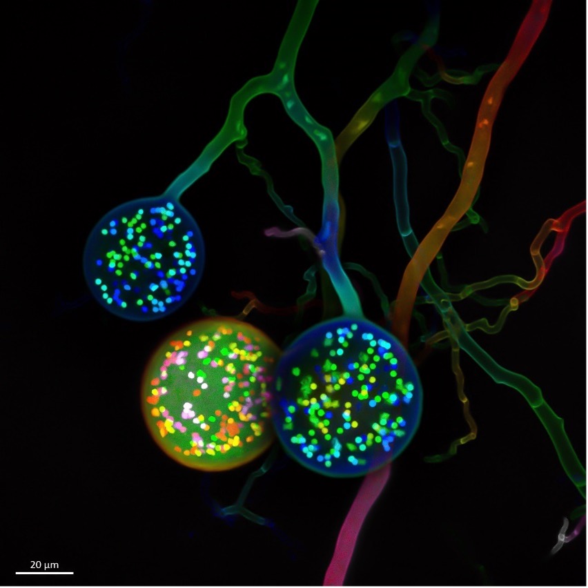
Image by Vasilis Kokkoris of multinucleate spores of a soil fungus. One cell typically carries one nucleus. In contrast, as seen here, an arbuscular mycorrhizal (AM) fungal cell carries hundreds of nuclei. This image was the 2021 winner for the EMEA region.
Q: What do you find most fascinating about microscopy?
Sian: There are so many incredibly powerful techniques in cell biology that can tell you all sorts of information, from the sequence of a gene to the phosphorylation status of a protein. But with microscopy, you can instantly see what is happening inside your sample. You also have the opportunity to see things that no other person has ever seen before.
Geoff: It opens up a whole world to everyone that we can’t see or appreciate without them. Finding macroscopic patterns or relationships is one of the best parts. Seeing the repetition of patterns and getting people to react and approach each image at an unconscious level. The best science engagement is often visual, and the microscope is a critical component of that.
Rachid: The output is an image, so it is very close to art.
Harini: Humans are visual creatures, and “seeing is believing.” My fascination with microscopy is twofold: it's a versatile quantitative research tool and a means to broadly disseminate imaging-based research to a lay audience. Microscopes have been indispensable research tools that have helped probe, capture, and understand many concepts of life science. Furthermore, the micrographs are an effective and engaging means of communicating science to both lay audience and the scientific community. The visual narrative transcends the barriers of language, education, expertise, and age making science accessible and understandable to all.
Q: What do you enjoy about evaluating the submissions for the Image of the Year Competition?
Stefan: The diversity and beauty of the imaged specimens.
Rachid: And the spectrum of applications.
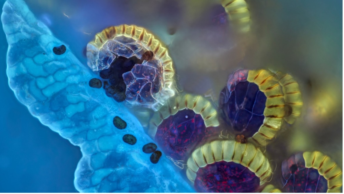
Colorful and intact fern sori spore capsules (sporangium) juxtaposed with an imploded one with spores bursting out. This is the 2021 IOTY winning image for Asia captured by Danial Han using Z-stacking.
Geoff: I love seeing the unique take that people have when selecting a submission for the competition. There is always something amazing and unexpected. It is both motivating and inspirational for my own work.
Sian: A lot of the images that I look at in my research are very similar to one another, as I mainly only use fluorescence microscopy. In the Image of the Year competition, I really enjoy seeing different types of microscopy images and I find the sheer diversity of microscopy techniques and images samples absolutely fascinating!
Harini: It's an honor to evaluate submissions from around the world. The best part of evaluating submissions is that you get to see beautiful micrographs of a wide variety of specimens and samples. It is truly a visual treat!
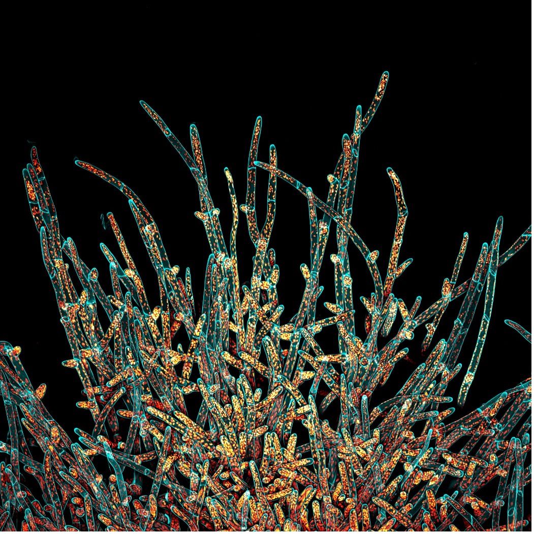
A maximum projection of the deconvolved Z-stack of moss Physcomitrium patens protonemal cells. The cell walls (in cyan) were stained live with calcofluor white, and the chloroplasts autofluorescence is in Fall LUT. Image captured by Ivan Radin, IOTY 2021 winner for the Americas.
Q: What qualities do you look for when evaluating the images?
Geoff: Composition and content. I look forward to seeing images with clear and defined statements, no distractions from cropping or debris or accidents. It doesn’t necessarily have to have the most complicated technique or staining but it really needs to have intelligently selected colors that are aesthetically pleasing with a complementary composition. The more pixels the better.
Sian: I look forward to seeing samples that I have never seen before—last year, I saw for the first time sea urchin muscles, moss, and Viagra crystals imaged with microscopes. There was even a sample that was imaged on a Pink Floyd CD case, which I can only dream of emulating in my own day-to-day research activities.
Harini: I look for a balance of good technique, stunning composition, and scientific relevance and narrative. It’s a balance between science and art. I hope to see some creative captioning along with the beautiful images to capture the audience.
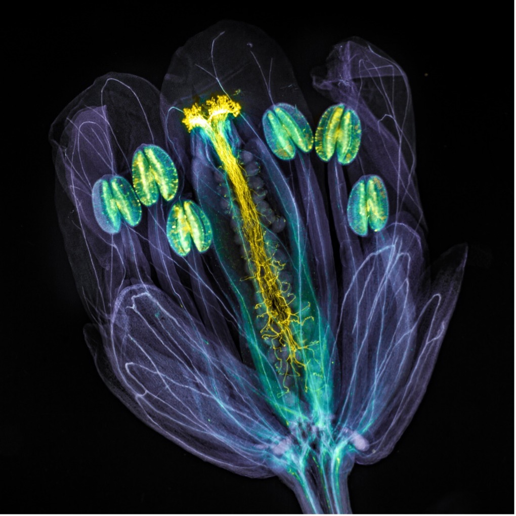
IOTY 2021 global winner Jan Martinek’s image of an Arabidopsis thaliana flower with pollen tubes growing through the pistil. The pollen tubes were stained with aniline blue (yellow fluorescence), while the flower tissues were chemically cleared to become transparent.
Q: Do you have any advice for IOTY award hopefuls?
Geoff: Pay attention to using the tool appropriately. Master the technique, understand how the photons travel and how to collect them most efficiently and represent what is there first. Once you have that then you can let that process become automatic, and it allows you to pay more attention to the content and composition and that in turn takes your scientific imaging to the next level and that generates more engagement for your science and is the best winning strategy for the IOTY competition.
Sian: I really like images where I notice different things every time I look at them. It’s great when the image has a striking focal point that your eyes are immediately drawn to, but also has lots of other interesting features that help the image to tell a story. I personally am also a big fan of colorful images!
Q: Any final comments?
Harini: I would like to congratulate the organizers and the participants for the success of the Image of the Year 2021 competition. I hope these images bring awareness and help to engage and entertain the general public about the wonders and significance of imaging science applied to life science research across scales: molecular, cellular, and tissue imaging.
Enter Up to 3 of Your Microscopy Images to IOTY 2022
If you are considering entering the IOTY 2022 competition, you have until February 27 at 10:00 p.m. EST (Feb. 28 at 12:00 p.m. JST) 2023. Upload 3 of your most captivating light microscopy images to increase your chances of capturing our jurors’ eyes and imaginations. Materials and life sciences subject matter are admissible.
If you need more convincing, go to the contest page and check out the prizes! We are excited to see this year’s submissions and wish you all the best.
Related Content
From Tinkerer to Prize Winner: Meet the 2021 Recipient of the IOTY Award for Asia
Celebrating the Global Image of the Year EMEA Winner’s Festive Fungi