Olympus Image of the Year Award 2021Image of the Year Award 2021 has come to a close. Have a look at below to see the beautiful images we received this year. For the latest information and submission details, click here! |
|

The Global Winner
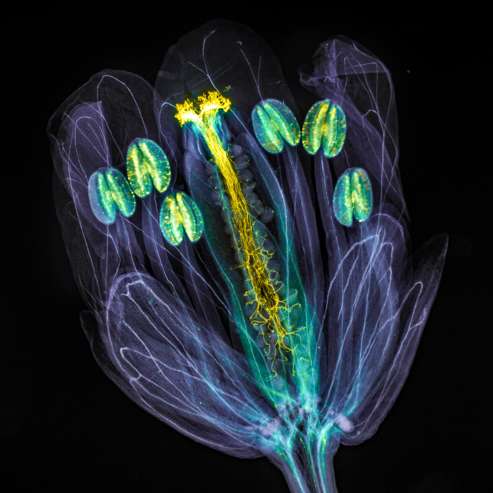 | The global winning image was taken by Jan Martinek (Czech Republic). Arabidopsis thaliana flower with pollen tubes growing through the pistil. The flower tissues were chemically cleared to become transparent, while the pollen tubes were stained with aniline blue (yellow fluorescence) in order to be seen. |
Regional Winners
Americas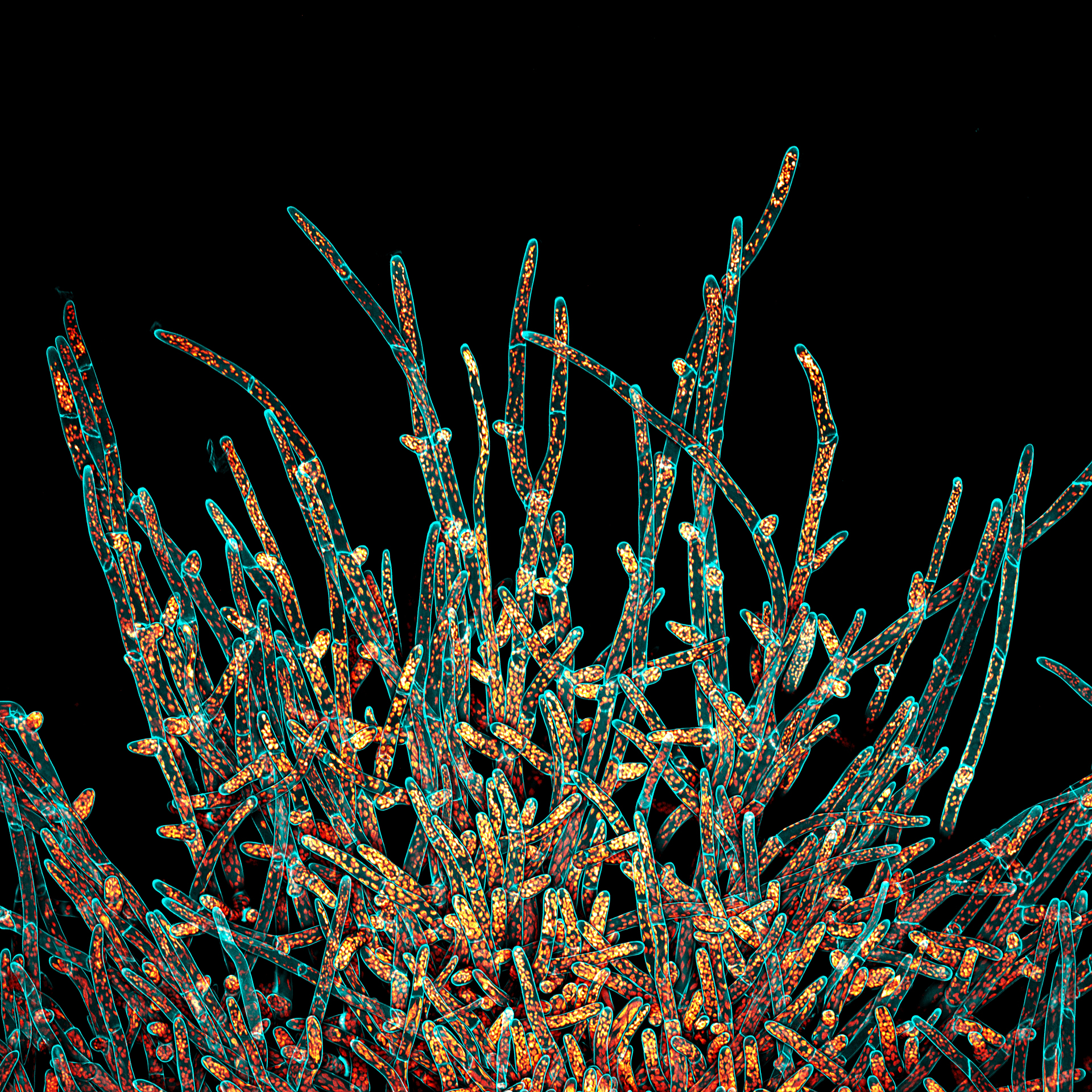 The winning image for the Americas was captured by Ivan Radin (USA). A maximum projection of the deconvolved Z-stack of moss Physcomitrium patens protonemal cells. Cell walls (in cyan) were stained live with calcofluor white. Chloroplasts autofluorescence is in Fall LUT. | Europe, the Middle East, and Africa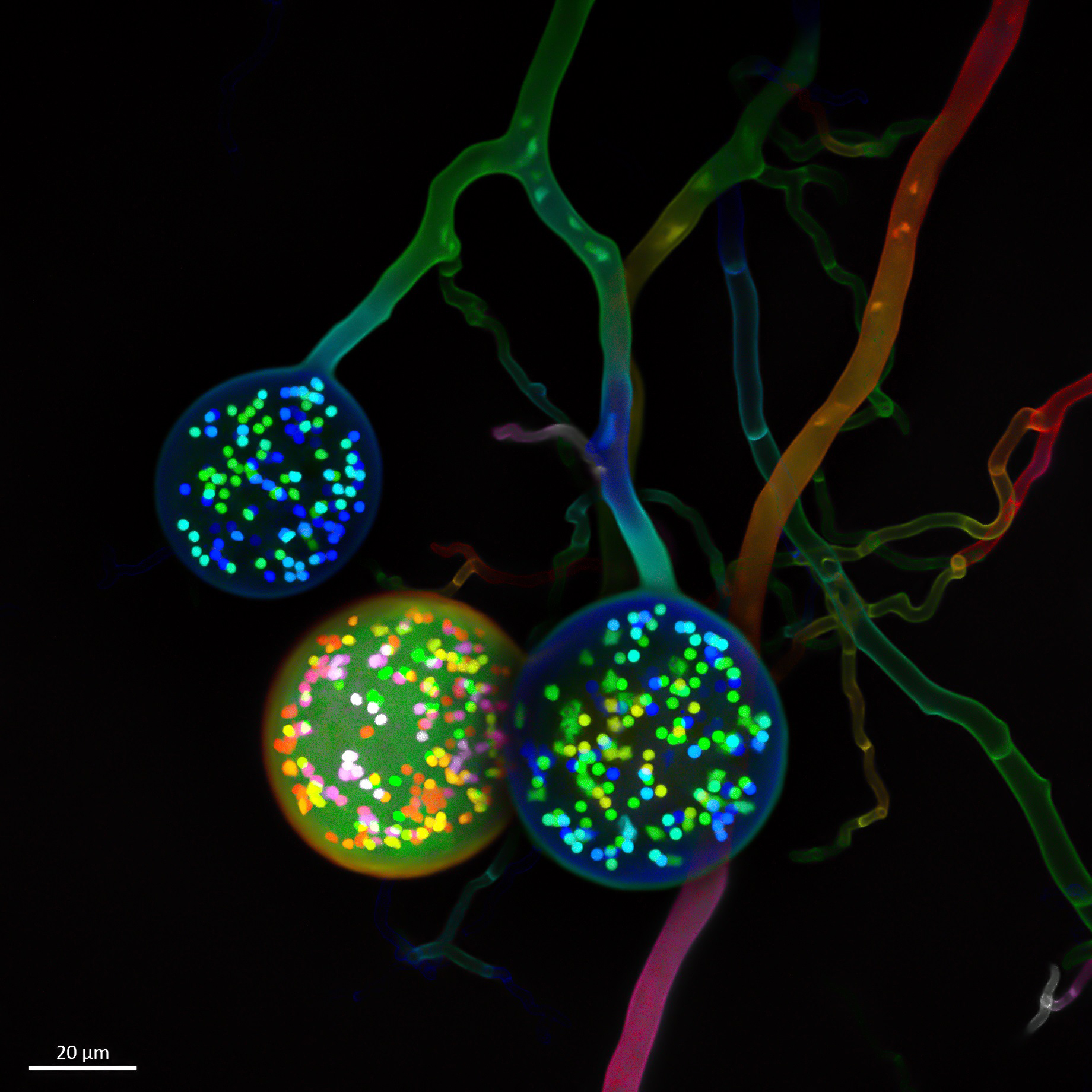 The winning image for EMEA was taken by Vasilis Kokkoris (Netherlands). Multinucleate spores of a soil fungus. One cell typically carries one nucleus. In contrast, as seen here, an arbuscular mycorrhizal (AM) fungal cell carries hundreds of nuclei. | Asia-Pacific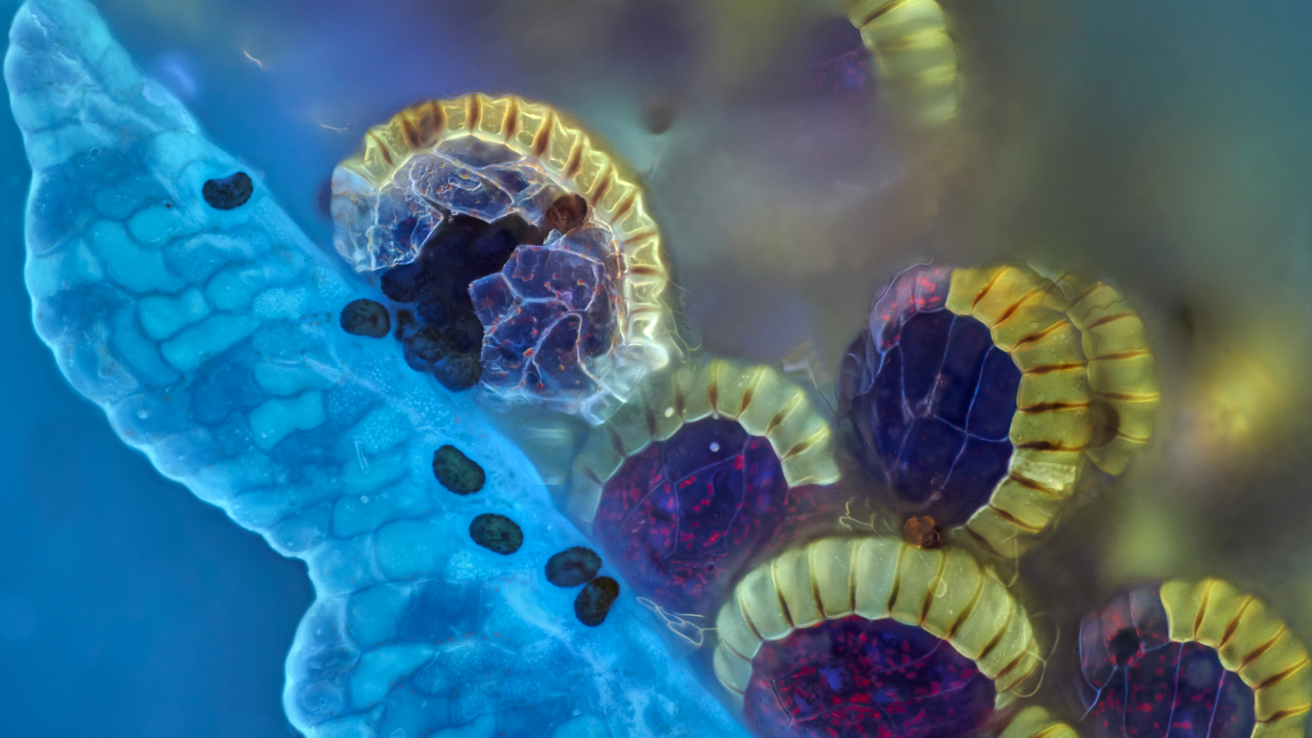 The winning image for Asia was captured by Daniel Han (Australia). Fern sori capsules with spores bursting out. Captured using Z-stacking. |
Honorable Mentions
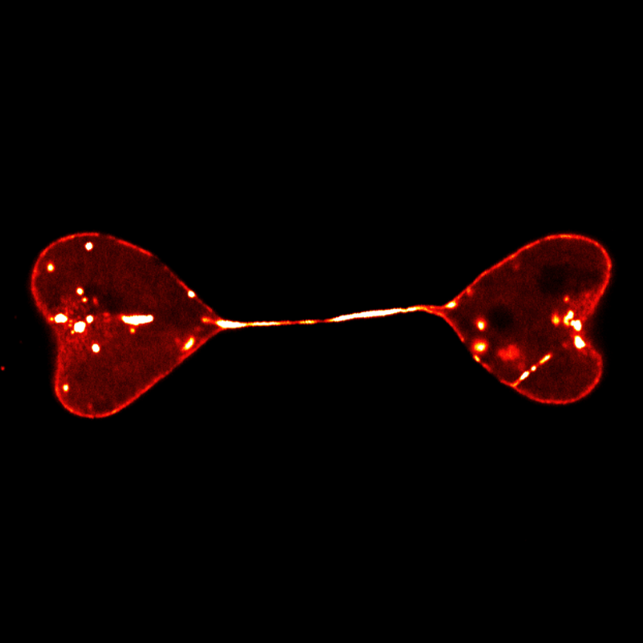 Semi-separated nuclei of two cells form a heart-to-heart shape. The nuclei were labeled by lamin. |  Ovaries of the fruit fly. | 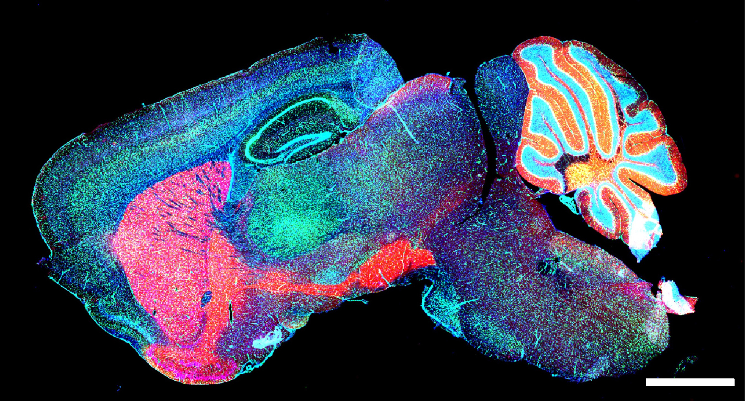 Mouse brain GABA neurons. |
 Autofluorescence image of Siberian polygala. Captured using confocal microscopy. Rendered using maximum projection. | 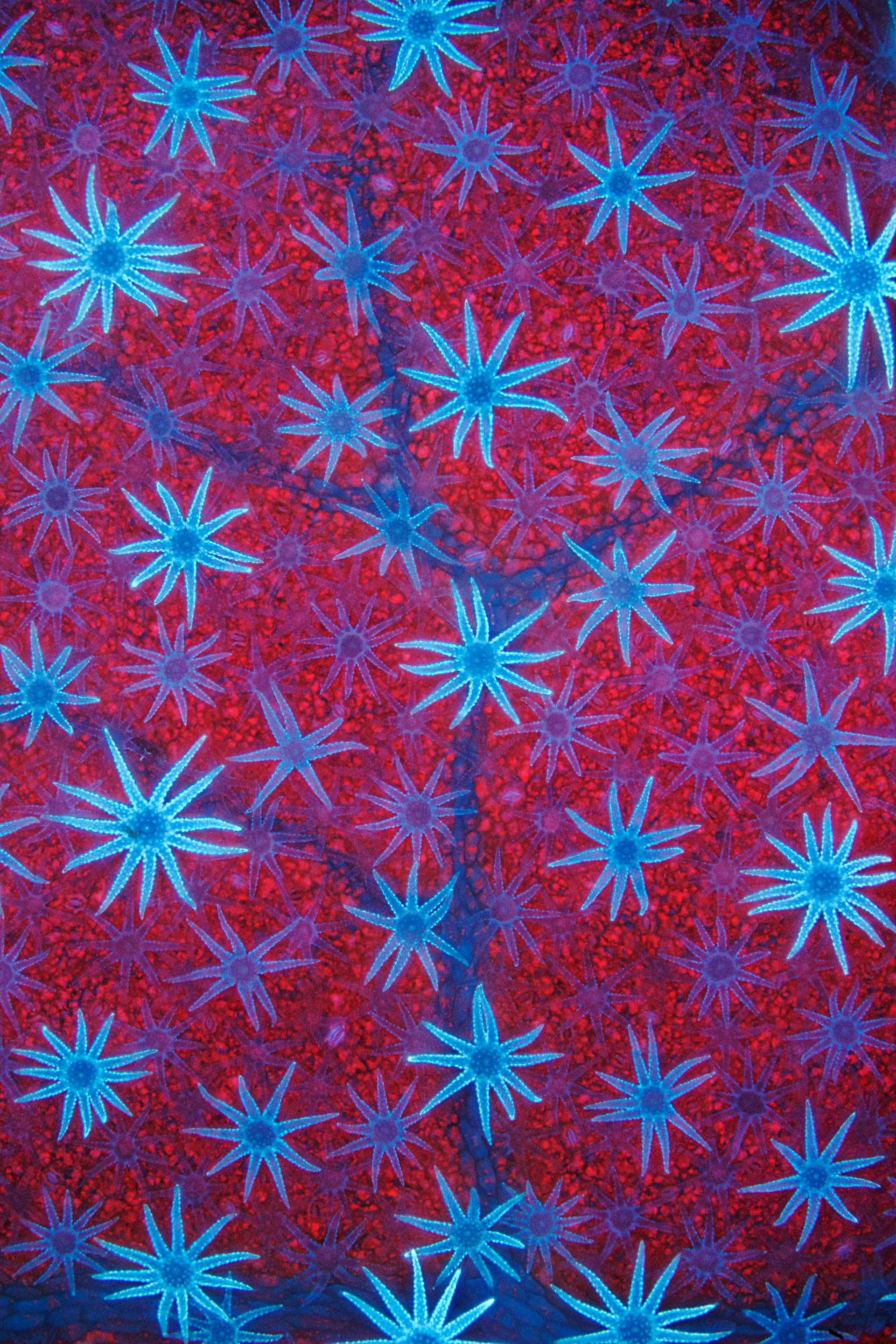 Blue autofluorescent, star-shaped defensive hairs cover the surface of a Deutzia leaf. The hairs are silhouetted against the leaf’s red-fluorescent, chlorophyll-packed cells. | 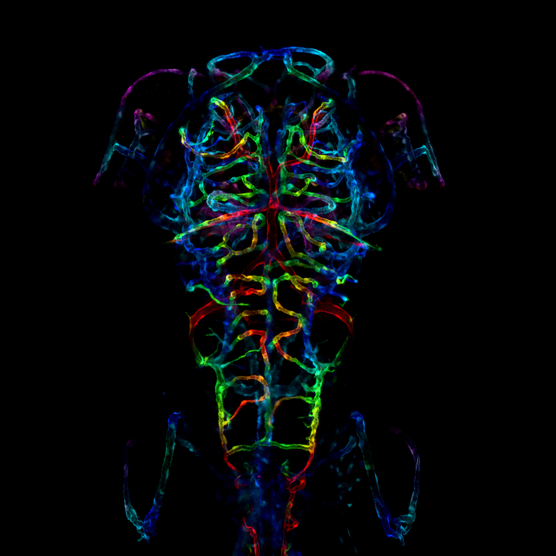 The developing nervous system of an embryonic zebrafish. Specifically, the image is a color-coded projection of the axonal projections of a zebrafish fixed six days after fertilization. |
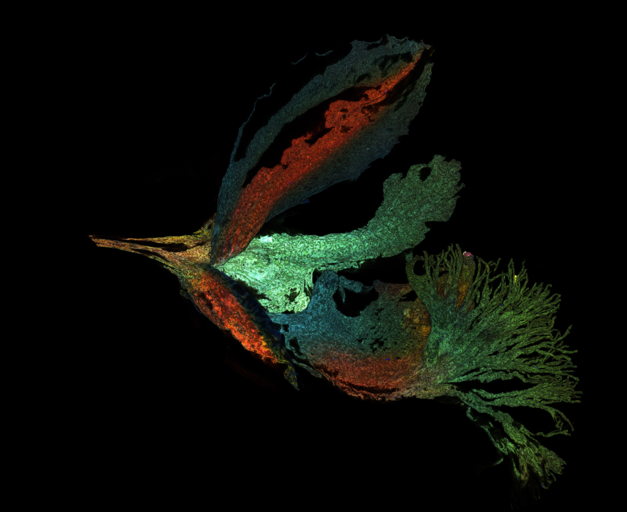
 Rasping tongue, or radula, of an Astraea conehead snail, Astraea tecta. Stained with Congo red. Imaged using a 10X (0.45 NA) objective. Depth color-coded projection. |
Download Wallpapers
Download the Image of the Year Award 2021 wallpaper package now for free and beautify your screen!
Download the wallpaper package for desktop (ZIP, 30.7 MB)
Download the wallpaper package for mobile (ZIP, 31.5 MB)
Virtual BackgroundsDownload your favorite virtual backgrounds and add them to your meetings! Download the virtual backgrounds package for your meeting application (jpg, 1.62 MB) | 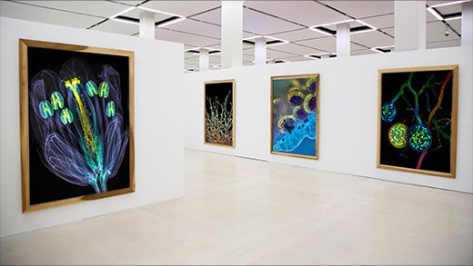 |
Sorry, this page is not
available in your country.
Sorry, this page is not
available in your country.