Evident LogoOlympus Logo

Ask the Experts

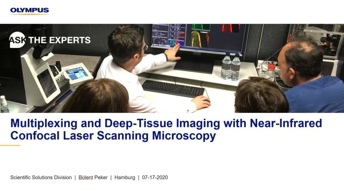
Multiplexing and Deep Tissue Imaging with Near-Infrared Confocal Laser Scanning Microscopy

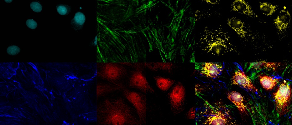
In this webinar, you’ll learn about fluorescence multiplexing and deep tissue imaging using near-infrared (NIR) laser light. NIR laser sources can help in visualizing biological structures more clearly and at higher resolution deep within a specimen. NIR excitation can also enable the use of more fluorescent dyes without spectral overlap.

Presenters:

Bülent Peker, Senior Product Marketing Manager
Rebecca Bonfig, Product Manager, Confocal Microscopy

FAQ

Webinar FAQs | Near-infrared (NIR) laser microscopy

Do you lose detection of lower wavelengths when using near infrared (NIR) imaging?

Due to the detectors’ design, you retain full availability to detect wavelengths starting at 400 nm. Lower wavelength emissions can still be collected at that range. We still have our standard and GaAsP photomultiplier tubes (PMTs) as well, so NIR is more like an add-on.

What are the benefits of using NIR over two-photon microscopy?

Two-photon microscopy is typically used for high-depth imaging in the red part of the spectrum. NIR offers an easy entry point into this application in a cost-effective way. With NIR, you can use confocal microscopes, so you don’t need to enter into a different class of microscopy system. As well as being much less expensive, NIR also offers greater multiplexing and spectral capabilities.

What is the VPH grating used for inside the microscope?

Volume-phase holographic (VPH) grating is part of our TruSpectral technology in standard and GaAsP detectors. It is used to generate the spectrum of light for spectral imaging. The advantage of VPH grating is that it is much more light efficient than using other reflection-type spectral gratings.

Do I have to purchase NIR lasers with the system or can I upgrade later?

One of the great features of this system is its modularity, so it can be upgraded at any time to add NIR lasers and detectors.

Is the six-detector mode available on both the IX and BX series?

Yes, the six-detector mode is available on both the IX series of inverted microscopes and the BX series of upright microscopes.

Does the FV3000 support NIR excitation wavelengths other than 730 and 785 nm?

The 730 nm and 785 nm laser lines are carefully selected as a result of cooperation with a specialized manufacturer that develops high-quality, stable lasers at these wavelengths.

However, there is a laser port available for the NIR range, so customization is possible. We encourage conversations to discuss the possibility of using different laser lines. The FV3000 microscope’s beam combiner makes it highly amenable to customization, and the system has optical coatings that support wavelengths out to 1600 nm, so users also have the ability to route their own NIR lasers into the system.

Can you image 730 and 785 nm on the same image set?

It is currently not possible to image both 730 nm and 785 nm, but we are always working to improve the system.

What are the differences between GaAsP detectors and competitors’ newest detectors?

We have not compared the systems side by side, but it’s important to remember that the detector is just one part of the whole system sensitivity. Make sure to look at the whole optical train and attached optics and how well they do in the NIR range. We know that there are several aspects during the light path that increase light efficiency in the NIR range, and it is also important that the optics are suited for the NIR range.

Can you use a white light laser with the FV3000 microscope?

White light lasers (WLLs) are not available on the FV3000 system. Olympus has a broad range of laser lines, which allow us to adapt the system for the needs of individual users and match the laser to their applications. There is also a cost-effectiveness factor and the advantage of the whole system not needing to be down if there is a problem with the WLL. Instead, you have individual laser lines that are affordable and easy to change.


관련 제품

컨포컬 레이저 스캐닝 현미경

FV4000

  • 매크로 규모에서 세포 내 구조까지 이미징을 위한 획기적인 동적 범위
  • TruSpectral 기술을 사용하여 최대 6개 채널을 동시에 멀티플렉싱
  • 고정 및 생세포 이미징을 위해 재설계된 고속, 고해상도 스캐너
  • 선도적인 NIR 기능과 유명한 광학 기술로 향상된 깊이와 감광도
  • 신뢰할 수 있고 반복 가능한 SilVIR 검출기를 안심하고 사용
  • 405nm~785nm의 더 넓은 스펙트럼 범위를 갖춘, 업계 최고 수준*의 10개 레이저 라인

*2023년 10월 기준.

Multiplexing and Deep Tissue Imaging with Near-Infrared Confocal Laser Scanning Microscopy

In this webinar, you’ll learn about fluorescence multiplexing and deep tissue imaging using near-infrared (NIR) laser light. NIR laser sources can help in visualizing biological structures more clearly and at higher resolution deep within a specimen. NIR excitation can also enable the use of more fluorescent dyes without spectral overlap.

FAQ

Webinar FAQs | Near-infrared (NIR) laser microscopy

Do you lose detection of lower wavelengths when using near infrared (NIR) imaging?

Due to the detectors’ design, you retain full availability to detect wavelengths starting at 400 nm. Lower wavelength emissions can still be collected at that range. We still have our standard and GaAsP photomultiplier tubes (PMTs) as well, so NIR is more like an add-on.

What are the benefits of using NIR over two-photon microscopy?

Two-photon microscopy is typically used for high-depth imaging in the red part of the spectrum. NIR offers an easy entry point into this application in a cost-effective way. With NIR, you can use confocal microscopes, so you don’t need to enter into a different class of microscopy system. As well as being much less expensive, NIR also offers greater multiplexing and spectral capabilities.

What is the VPH grating used for inside the microscope?

Volume-phase holographic (VPH) grating is part of our TruSpectral technology in standard and GaAsP detectors. It is used to generate the spectrum of light for spectral imaging. The advantage of VPH grating is that it is much more light efficient than using other reflection-type spectral gratings.

Do I have to purchase NIR lasers with the system or can I upgrade later?

One of the great features of this system is its modularity, so it can be upgraded at any time to add NIR lasers and detectors.

Is the six-detector mode available on both the IX and BX series?

Yes, the six-detector mode is available on both the IX series of inverted microscopes and the BX series of upright microscopes.

Does the FV3000 support NIR excitation wavelengths other than 730 and 785 nm?

The 730 nm and 785 nm laser lines are carefully selected as a result of cooperation with a specialized manufacturer that develops high-quality, stable lasers at these wavelengths.

However, there is a laser port available for the NIR range, so customization is possible. We encourage conversations to discuss the possibility of using different laser lines. The FV3000 microscope’s beam combiner makes it highly amenable to customization, and the system has optical coatings that support wavelengths out to 1600 nm, so users also have the ability to route their own NIR lasers into the system.

Can you image 730 and 785 nm on the same image set?

It is currently not possible to image both 730 nm and 785 nm, but we are always working to improve the system.

What are the differences between GaAsP detectors and competitors’ newest detectors?

We have not compared the systems side by side, but it’s important to remember that the detector is just one part of the whole system sensitivity. Make sure to look at the whole optical train and attached optics and how well they do in the NIR range. We know that there are several aspects during the light path that increase light efficiency in the NIR range, and it is also important that the optics are suited for the NIR range.

Can you use a white light laser with the FV3000 microscope?

White light lasers (WLLs) are not available on the FV3000 system. Olympus has a broad range of laser lines, which allow us to adapt the system for the needs of individual users and match the laser to their applications. There is also a cost-effectiveness factor and the advantage of the whole system not needing to be down if there is a problem with the WLL. Instead, you have individual laser lines that are affordable and easy to change.


관련 제품

컨포컬 레이저 스캐닝 현미경

FV4000

  • 매크로 규모에서 세포 내 구조까지 이미징을 위한 획기적인 동적 범위
  • TruSpectral 기술을 사용하여 최대 6개 채널을 동시에 멀티플렉싱
  • 고정 및 생세포 이미징을 위해 재설계된 고속, 고해상도 스캐너
  • 선도적인 NIR 기능과 유명한 광학 기술로 향상된 깊이와 감광도
  • 신뢰할 수 있고 반복 가능한 SilVIR 검출기를 안심하고 사용
  • 405nm~785nm의 더 넓은 스펙트럼 범위를 갖춘, 업계 최고 수준*의 10개 레이저 라인

*2023년 10월 기준.

Experts
Bülent Peker
제품 마케팅 수석 관리자
Evident

안녕하세요. Bülent Peker 입니다. 레이저 스캐닝 현미경의 지정 전문가입니다. 물리화학 박사 과정에서 시간 분해 2광자 현미경을 연구하였으며 이때 현미경 및 광산업에 처음으로 관심을 가지게 되었는데 이후 이러한 열정이 계속 되었습니다. Olympus에 일한 지 13년이 넘었으며 첨단 레이저 스캐닝 현미경 시판 팀에서 활동하였습니다. 다광자 시스템의 응용과 레이저 스캐닝 시스템의 맞춤화 가능성에 특히 관심이 많습니다. 

Rebecca Bonfig
제품 관리자, 공초점 현미경

안녕하세요. Olympus의 공초점 현미경 제품 관리자인 Rebecca Bonfig입니다. 루이빌 대학에서 물리학 및 생물물리학과 대학원 과정을 이수하고 부갑상선 호르몬에 의한 신장 인산염 운반기 Npt2a의 전사 후 조절을 연구하였습니다. 2015년에 Olympus에 입사하였으며 현재 미국, 캐나다 및 라틴 아메리카의 FLUOVIEW 제품 라인을 지원하고 있습니다.

Multiplexing and Deep Tissue Imaging with Near-Infrared Confocal Laser Scanning Microscopy2024년12월15일
죄송합니다. 이 페이지는 해당 국가에서 사용할 수 없습니다.
Ask the Expert Sign-up

By clicking subscribe you are agreeing to our privacy policy which can be found here.

Sorry, this page is not
available in your country.

죄송합니다. 이 페이지는 해당 국가에서 사용할 수 없습니다.