Last updated on September 13, 2024.
Applying more than 100 years of optical development history and knowledge gained from communicating with researchers, we strive to support researchers’ imaging workflows with cutting-edge solutions. We continuously seek ways to leverage our expertise to solve issues with your experiments. To demonstrate some ways we do this, I’ll present a case study, an application, and four workflow solutions that improve the efficiency of experiments—from the cell culture stage to imaging and image analysis.
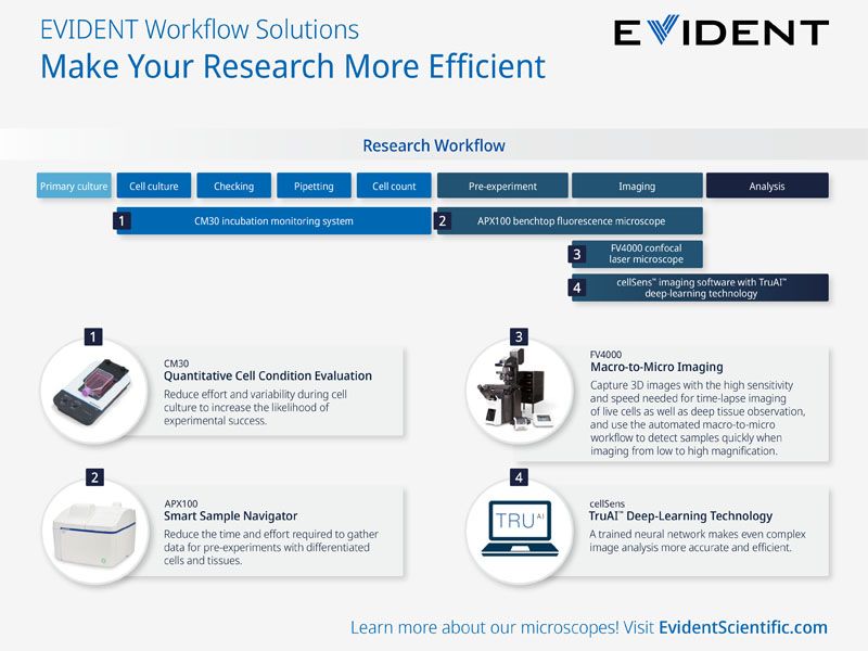
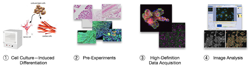
Case Study: Standardizing Cell-Preparation Stages for More Efficient Subsequent Experiments
When conducting experiments involving differentiation induction from stem cells, you can use our solutions to optimize efficiency during each of the following four steps:
- Quantitatively evaluating cell culture conditions using the CM30 incubation monitoring system increases the likelihood that cells will form the targeted tissues, making subsequent experiments more efficient.
Read this whitepaper on the key to achieving a successful cell culture workflow. - In pre-experiments with differentiated cells and tissues, you can acquire more high-quality data in less time and with less effort using the APEXVIEW™ APX100 benchtop fluorescence microscope.
Read this blog post to learn how the APX100 microscope simplifies pre-experiment steps. - At the data acquisition step, confocal imaging is performed to evaluate the function of the sample. Using the FLUOVIEW™ FV4000 confocal laser scanning microscope, it’s possible to acquire high-definition images of the region of interest while capturing the entire sample.
- Using the TruAI™ deep-learning module of cellSens™ imaging software enables high-precision image analysis.
With this set of four workflow improvements, you can expect to achieve more efficient and effective results.
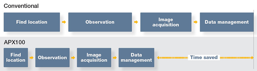
Comparing EVIDENT Workflow Solutions with Conventional Methods
As you can see in the table below, many of the cell culture workflow tasks that traditionally had to be performed manually can be automated utilizing AI and modern tools, saving time. Even if an individual task is not especially time consuming, it is often repeated numerous times for different cell types, which means that the time savings over the long term can be significant.
And the benefits aren't just about efficiency. The quality of experiments can also be improved through factors such as reducing the burden on cells and preventing the risk for human error due to manual manipulations.
| Conventional methods | Evident workflow solutions | ||
|---|---|---|---|
| Cell culture | Cell check method | Necessary to take the culture out of the incubator each time | Remote check available, avoid removing the culture from the incubator |
| Cell count and density measurement | Conducted with cell counting equipment, etc. | Automatic measurement while monitoring | |
| Culture conditions | Varies from person to person | Quantitative | |
| Pre-experiment | Finding the observation position |
Visual and microscope operation
|
Autodetection
|
| Data acquisition | Multiple-specimen imaging | One sample at a time | Automatic imaging of multiple samples |
| Image analysis | Detection of specific specimens | Manually locate a specific specimen | Autodetection using AI |
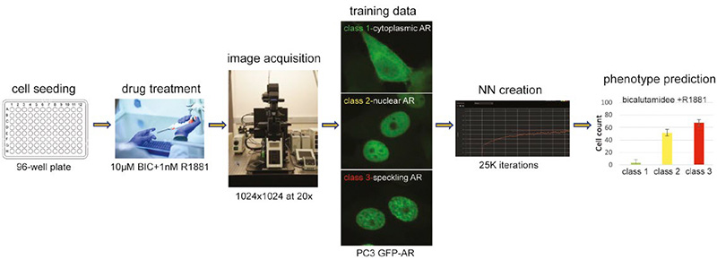
Application: Predicting Multi-Class Nuclei Phenotypes for Drug Testing Using Deep Learning
In drug discovery research, trained neural networks (NNs) can be applied to accelerate phenotype prediction. Using the FV4000 confocal laser scanning microscope and the TruAI deep-learning technology of cellSens imaging software, researchers can estimate nuclear positions and segment them without using nuclear staining, and then construct a method to classify cells in one step based on changes in AR phenotype according to the drug. This approach reduces the time and cost of experimentation and contributes to the efficiency of chemical testing.
Learn more about this application
Products Used for These Workflow Solutions
Meet the products that contribute to the workflow solutions introduced in this blog post.
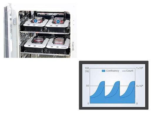
CM30 Incubation Monitoring System
|
|
APEXVIEW APX100 Benchtop Fluorescence MicroscopeThe APX100 system's smart sample navigator automatically acquires a macro image and the built-in AI detects your sample on the slide. The system’s simple and intuitive software and its macro image acquisition feature provides a stress-free experiment process, so obtaining high-quality images is easy for all users. | Related Videos |
 | |
FLUOVIEW FV4000 Confocal Laser Scanning MicroscopePinpointing the area of interest in your sample can be time-consuming. The FV4000 system makes it easy to acquire stitched images from low magnification (1.25x or 2x) to high magnification. By connecting macro low-magnification images with micro high-resolution images, the FV4000 system not only provides continuous macro-to-micro observation, but also automates it for the user. Thanks to the SilVIR™ detector and the Microscope Performance Monitor (MPM), the acquired images offer reliable quantitative data for analysis. | 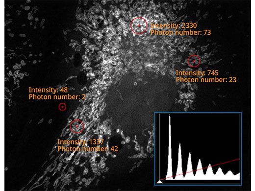 |
cellSens Imaging SoftwareOur cellSens imaging software facilitates various image analysis tasks, such as luminance analysis, object measurement and classification, and tracking analysis on image data acquired with a microscope. Using the TruAI deep-learning module for object detection enables automatic analysis of images that were manually analyzed in the past, so results are obtained more efficiently and with improved accuracy. | |
 Blue: Detecting the nuclei with high accuracy despite scratches and dust on the vessel. | |
Related Content
Streamline Your Research Workflow with a Benchtop Fluorescence Microscope
Drug Viability Testing of 3D Cancer Spheroids Using Automated Macro-to-Micro Imaging
Label-Free Quantification You Can Count On: A Deep Learning Experiment