Rotifers, also known as wheel animals, are microscopic aquatic animals belonging to the phylum Rotifera. They get their name from the ciliated crowns located on their head, a characteristic structure used for both locomotion and gathering food particles.
The corona is made of beating cilia, which give the impression of rotating wheels—thus where their name comes from!
With over 2,000 different species, rotifers can be found all over the world. Rotifers live mostly in freshwater habitats such as ponds and lakes. You can also find rotifers in mosses, lichens, soils, sewages, marine environments, permafrost, and even inside or outside other animals.

Collecting rotifer specimens to view under the microscope. Image courtesy of Chloé Savard.
Although a few rotifer species are parasitic, most rotifers are free-living and either swim amongst plankton, crawl within sediments, attach themselves to submerged aquatic plants, or live inside a secreted, gelatinous tube. These tiny invertebrates form an extremely diverse group that developed many different morphologies and locomotion strategies to adapt to various environmental pressures, including predation.
General Biology of Rotifers
Are rotifers unicellular or multicellular? At first sight, rotifers may look like unicellular microorganisms. They’re actually made of around 1,000 cells, despite being smaller than a lot of unicellular protozoans.
Rotifers can reach up to 2 mm but usually measure 0.1 to 0.5 mm long. The smallest rotifers that exist are only six times the size of a human red blood cell, which is around 0.006 to 0.008 mm (6–8 µm).
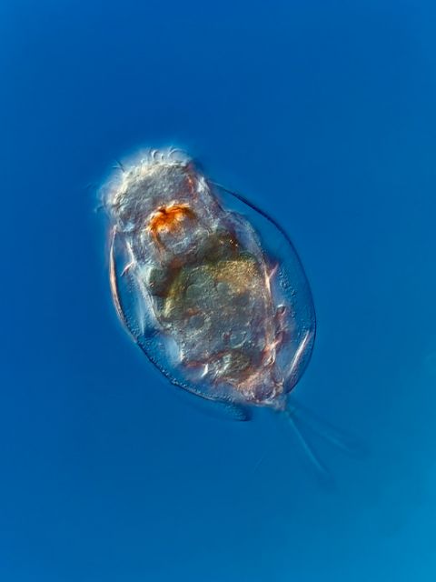
Euchlanis sp. rotifer captured using differential interference contrast (DIC) microscopy. Image courtesy of Chloé Savard.
Rotifers exhibit a wide range of morphologies, but they share the same main body regions: a head (corona), a body (trunk), and a foot. The foot, when present, can possess separated toes—usually two. Some rotifer species can have up to four toes, while others don’t have any toes at all.
Depending on the species, the foot can extend telescopically. This phenomenon can especially be observed within bdelloid rotifers but also within the Rotaria neptuna species. The foot may also possess pedal glands, which secrete a sticky substance. Rotifers use this substance as an adhesive to temporarily attach to different substrates, such as plants, rocks, pieces of wood, and animals.
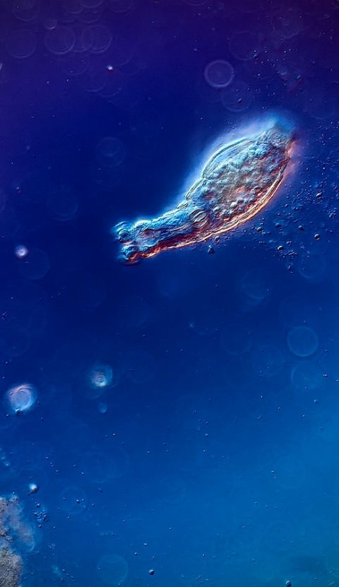
Bdelloid rotifer captured using DIC microscopy at 200X. Image courtesy of Chloé Savard.
In most planktonic and free-swimming rotifer species, the foot is absent. Some rotifers are strictly sessile types; they can swim around in their larval stage until they find the perfect substrate to attach themselves for the rest of their lives. This is the case of Stephanoceros fimbriatus.
Even though these primitive creatures don’t possess either a circulatory or respiratory system, rotifers do have a brain, small red eyes, a complete digestive tract, and a muscular, reproductive, and excretory system.
To protect most of the organ systems located inside the trunk, rotifers possess a body wall, also called the integument, which can be either very thick and hard or thin and flexible. Those who possess a hard shell are known as loricate rotifers, and those who are flexible are referred to as illoricated rotifers.
In some loricate rotifers, the integument can bear spines at the anterior or posterior end that serve as a defense against predators. It has been shown in certain Keratella and Asplanchna species that spines can be produced in response to soluble chemical signals released by invertebrate predators such as copepods. The more predators present in Keratella’s environment, the more chemical signals are present and the more these rotifers are inclined to develop spines.
The head of rotifers bears a corona with two ciliated rings that create a water vortex used to gather food particles but also to swim around in the water column.
Like tardigrades, bdelloid rotifers have the ability to enter a dormant state called cryptobiosis, or more specifically, anhydrobiosis. In this state, they can survive desiccation, meaning the complete loss of water from their body, for long periods of time.
Bdelloid rotifers can enter anhydrobiosis to escape fungal parasites or to survive extreme cold or exposure to ionizing radiation. Fun fact: in a study from June 2021, a bdelloid rotifer retrieved from the Arctic’s permafrost came out of anhydrobiosis after being frozen for 24,000 years!
What Do Rotifers Eat?
Alongside microcrustaceans and protists, rotifers form the third group that dominates freshwater zooplankton. With a diet consisting of organic detritus, bacteria, yeasts, algae, small ciliates, and other protozoans, rotifers can adapt their feeding behaviour depending on environmental conditions and prey abundance.
Some rotifers are even large enough to prey on smaller rotifers!
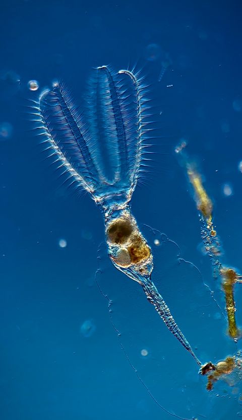
Stephanoceros fimbriatus rotifer captured using DIC microscopy at 100X. Image courtesy of Chloé Savard.
Using their ciliated corona, rotifers create a water vortex that traps food particles, bringing them directly to the mouth. After entering the mouth, food is crushed by a modified pharynx called the mastax—a unique characteristic to rotifers. When chewing food, the mastax looks a bit like a beating heart, which can be confusing at first since rotifers do not possess any circulatory system.
The muscular mastax has a jaw-like structure called the trophi, which helps to grasp, grind, and pierce various food material. The trophi’s composition is different from one species to another, so it is a valuable tool when trying to identify rotifers. After being crushed, food exits the mastax and goes down the esophagus until it reaches the stomach, the intestines, and ultimately the anus where it exits the body.
Rotifers can also be preyed upon themselves by many aquatic predators such as protists (especially ciliates), insects, other rotifers, cladocerans, copepods, and fish. To escape predators, rotifers have developed many strategies: jumping movements, mucus sheaths, spines, and thickened shells.
How Do Rotifers Reproduce?
Rotifer reproduction methods vary enormously within the rotifer phylum. Rotifers either reproduce sexually or asexually. In both cases, they need to conceive eggs.
In rotifer species that reproduce sexually, both males and females can be observed in the population. Female rotifers produce mictic eggs, which are haploid and need to be fertilized by a male’s spermatozoid. If these eggs aren’t fertilized, they will develop parthenogenetically into males. In planktonic species reproducing sexually, sexual dimorphism can often be observed; male rotifers don’t possess a foot, are smaller, and swim faster than female rotifers.
On the opposite side of the spectrum, bdelloid rotifers reproduce asexually by parthenogenesis. Their populations are composed of females only. Further, eggs don’t need to be fertilized by a male’s spermatozoid to give rise to female baby rotifers. These types of eggs are called amictic and are diploid.
Bdelloid rotifers are one of the few animal groups that lack male representatives alongside certain species of cladocerans, aphids, bees, and ants. It has been roughly 40 million years that bdelloid rotifers have been evolving as an all-female group!
How Do You View Rotifers Under a Microscope?
Rotifers can be found almost everywhere. By taking samples either from a pond, lake, or even from your building’s gutters, you can be certain to find at least one species of rotifer to view under the microscope.

Collecting rotifer specimens to view under the microscope. Images courtesy of Chloé Savard.
Bdelloid rotifers are especially abundant in mosses and lichens. Since they are likely in their dormant state, it’s important to soak the moss or lichen for 24 hours before squeezing the water out of it and placing the petri dish under the microscope.
Bdelloid rotifers often crawl instead of swim, so they can be mistaken for a worm if you haven’t seen any before (just like I did the first time I saw one!) For optimal observations of rotifers, use brightfield or darkfield illumination on a compound microscope.
References and Further Reading
Allen, A. A. (1968). Morphology of the planktonic rotifer Polyarthra vulgaris. Transactions of the American Microscopical Society, 60-69.
Bogdan, K. G., & Gilbert, J. J. (1982). Seasonal patterns of feeding by natural populations of Keratella, Polyarthra, and Bosmina: Clearance rates, selectivities, and contributions to community grazing 1. Limnology and Oceanography, 27(5), 918-934.
De Paggi, S. B. J., Wallace, R., Fontaneto, D., & Marinone, M. C. (2020). Phylum Rotifera. Thorp and Covich’s Freshwater Invertebrates, 145–200.
Gladyshev, E., & Meselson, M. (2008). Extreme resistance of bdelloid rotifers to ionizing radiation. Proceedings of the National Academy of Sciences, 105(13), 5139-5144.
Gribble, K. E., & Snell, T. W. (2018). Rotifers as a Model for the Biology of Aging. Conn’s Handbook of Models for Human Aging, 483–495.
Hickman, C. P., Roberts, L. S., Larson, A., Ober, W. C., & Garrison, C. (2015). Animal
diversity. WC Brown. 183.
Ricci, C. (1998). Anhydrobiotic capabilities of bdelloid rotifers. Hydrobiologia, 387, 321-326.
Segers, H., & De Smet, W. H. (2007). Diversity and endemism in Rotifera: a review, and Keratella Bory de St Vincent. Protist Diversity and Geographical Distribution, 69-82.
Shmakova, L., Malavin, S., Iakovenko, N., Vishnivetskaya, T., Shain, D., Plewka, M., & Rivkina, E. (2021). A living bdelloid rotifer from 24,000-year-old Arctic permafrost. Current Biology, 31(11), R712-R713.
Taylor, R. A. J. (2019). Other invertebrates. Taylor’s Power Law, 305–326.
Wallace, R. L. (2002). Rotifers: exquisite metazoans. Integrative and Comparative Biology, 42(3), 660-667.
Wallace, R. L., Snell, T. W., Walsh, E. J., Sarma, S. S. S., & Segers, H. (2019). Phylum Rotifera. Thorp and Covich’s Freshwater Invertebrates, 219–267.
Welch, D. B. M., & Meselson, M. (2000). Evidence for the evolution of bdelloid rotifers without sexual reproduction or genetic exchange. Science, 288(5469), 1211-1215.
Wininger, J. D. (2004). Parthenogenetic Stem Cells. Handbook of Stem Cells, 635–637.
Related Content
Avalon Finds Her Dream Microscope to Study Rotifers
Stars of the Show—Our Most Popular Microscope Images for July 2023
Easy Being Green—Our Most Popular Microscope Images for June 2023