Not Available in Your Country
Sorry, this page is not
available in your country.
Overview
 | Flexible and CustomizableThe LED illuminator for the BX53 is equivalent to or better than a 100 W halogen lamp, delivering brightness that’s appropriate for teaching or contrast methods. The BX53 microscope can be customized for different observation methods, such as phase contrast and fluorescence, with modular components. |
|---|
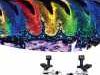
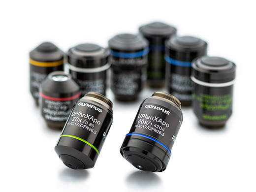
Acquire Precise Images with X Line ObjectivesThese objectives combine improved flatness, numerical aperture, and chromatic aberration to deliver clear, high-resolution images with better color accuracy across the entire spectrum. The elimination of violet color aberration creates clear whites and vivid pinks, improving contrast and sharpness. |
|
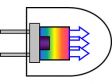
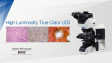
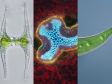
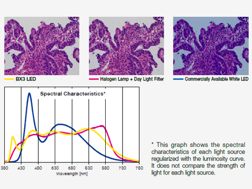 | Bright LED Lighting Designed for Pathology and LaboratoryWith spectral characteristics similar to halogen light sources, the LED illuminator enables you to view the purple, cyan, and pink colors important in clinical research applications while enjoying the long use life of an LED. |
|---|
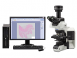
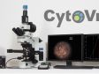
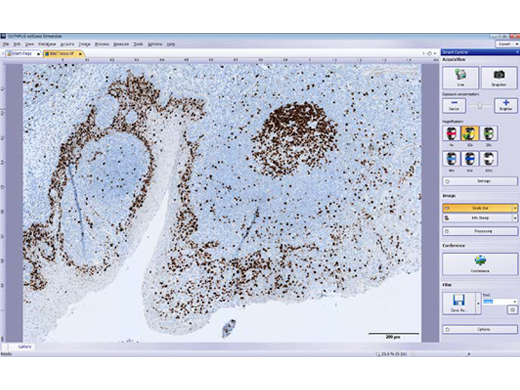
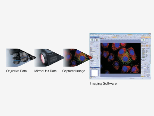
Digital Imaging Helps Simplify Your WorkFrom advanced research to capturing images for conferences, our digital microscope cameras and cellSens imaging software help ensure fluorescence imaging with a high signal-to-noise ratio. |
|
|---|
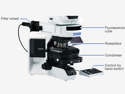
| Upgrade to Motorized ComponentsAutomatically record and share magnification setting information with the optional coded nosepiece or change observation methods with a single touch using the hand switch. Motorized options include:
|
|---|
Bright Images in Multi-Head ConfigurationsThe multi-head light path has been redesigned not only to orient all images the same way but also to allow the intense LED to provide clear, bright images for up to 26 participants. | 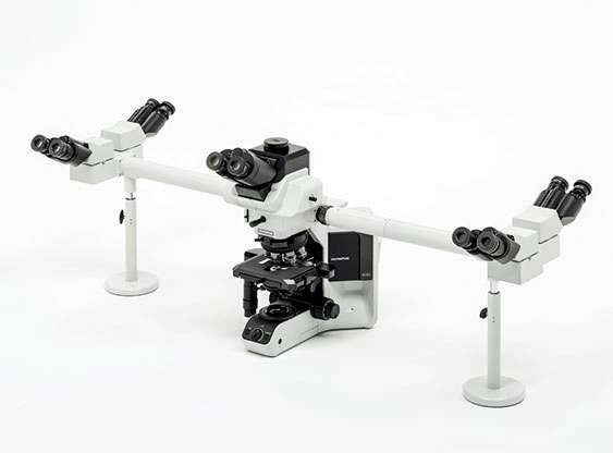 |
|---|
| Save Microscopy Data via Coded UnitsAdd an optional coded nosepiece to your BX53 microscope to automatically record and share magnification setting information for post-imaging treatments. The metadata is automatically sent to cellSens software, helping minimize mistakes and scaling errors |
|---|
Need assistance? |
Applied Technologies
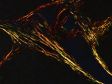
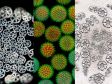
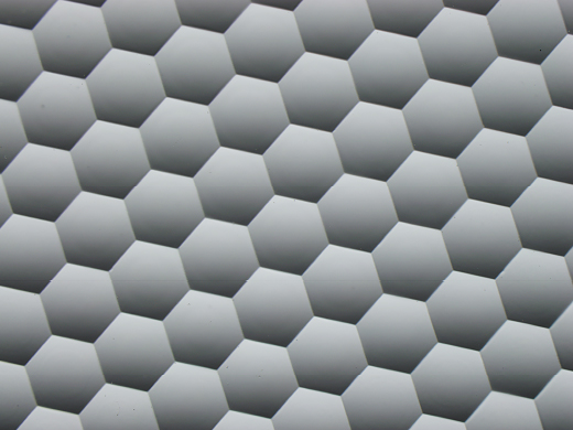
Surface of Fly-eye Lens, Enlarged Image | Even Fluorescence Across the Field of ViewFluorescence illuminators with a fly-eye lens provide homogenous illumination across the entire wavelength spectrum and simplify burner alignment. |
|---|
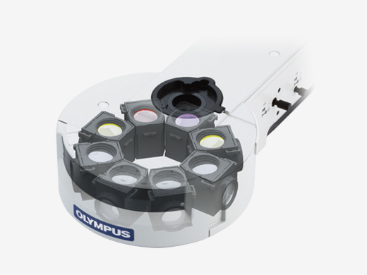
Flexible 8-Position IlluminatorEasily changeable mirror units to provide flexibility for a wide variety of fluorescence specimens. The design reduces the need to replace mirror units for multicolor or FISH applications, further accelerating your work. |
|
|---|
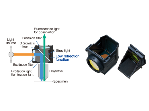
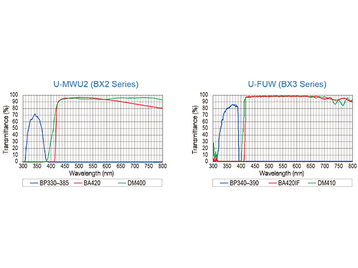
Mirror Units Optimized for FluorescenceThe fluorescence mirror units have advanced coatings that provide high transmission and steep cut-off slopes. The interior surfaces eliminate over 99% of stray light for high sensitivity and sharp color separation. |
 |  |
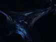
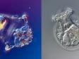
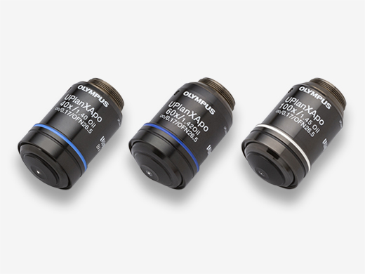
| High-Transmission Objectives with Reduced AutofluorescenceUIS2 objectives with a high numerical aperture (NA) have chromatic aberration corrections to provide high resolution, even from faint fluorescence signals. Advanced coating technologies reduce autofluorescence and improve the signal-to-noise ratio for flat, high transmission over a wide wavelength. |
|---|
Condenser Reduces Back ReflectionsDuring fluorescence imaging, the motorized universal condenser reduces back reflections and autofluorescence by swinging its top lens out, automatically closing its diaphragm to a minimum, and locating the wheel between two positions. |
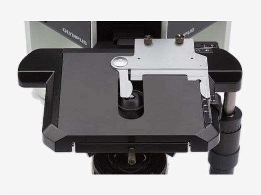
| Comfortable StageMechanical stages are coated with an abrasion-resistant ceramic and have a rackless, wire-driven design. |
|---|
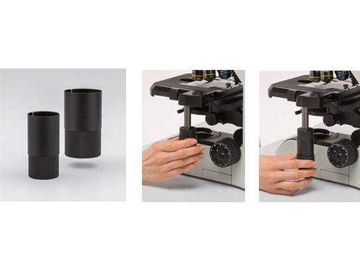
Keep Your Arms Resting on the DeskThe stage handle extender enables you to keep your arms resting on the desk while you work. A rubber cap can be mounted to the handle, so you can control the stage with small force. |
|
|---|
Need assistance? |
Specifications
| Observation Method > Brightfield | ✓ | |
|---|---|---|
| Observation Method > Darkfield | ✓ | |
| Observation Method > Phase Contrast | ✓ | |
| Observation Method > Fluorescence (Blue/Green Excitations) | ✓ | |
| Observation Method > Fluorescence (Ultraviolet Excitations) | ✓ | |
| Observation Method > Differential Interference Contrast | ✓ | |
| Observation Method > Polarized Light | ✓ | |
| Observation Method > Simple Polarized Light | ✓ | |
| Illuminator > Transmitted Köhler Illuminator > LED Lamp | ✓ | |
| Illuminator > Fluorescence Illuminator > 100 W Mercury Lamp | ✓ | |
| Illuminator > Fluorescence Illuminator > Light Guide Illumination | ✓ | |
| Focus > Focusing Mechanism > Stage Focus | ✓ | |
| Intermediate Magnification Changer > Manual Terret | ✓ | |
| Revolving Nosepiece > Motorized (7 positions) | ✓ | |
| Revolving Nosepiece > Manual > Coded (7 positions) | ✓ | |
| Revolving Nosepiece > Manual > Standard (7 positions) | ✓ | |
| Stage > Manual > Manual Stages with Right-Hand Control |
| |
| Stage > Mechanical > Oil Rectangular Stage with Right-Hand Control |
| |
| Stage > Mechanical > Plain Stage | Dimensions: 180 mm × 150 mm (7 in. × 6 in.) | |
| Stage > Mechanical > Rotatable Graduated Stage |
| |
| Condenser > Motorized > Universal Condenser | NA 0.9/ W.D. 1.5 mm for 1.25X–100X [swing-out: 1.25X–4X, with oil top lens: (NA 1.4/ W.D. 0.63 mm)] | |
| Condenser > Manual > Universal Condenser | NA 0.9/ W.D. 1.5 mm for 1.25X–100X [swing-out: 1.25X–4X, with oil top lens: (NA 1.4/ W.D. 0.63 mm)] | |
| Condenser > Manual > Swing-Out Condenser | NA 0.9/ W.D. 2 mm (1.25X–100X) | |
| Condenser > Manual > Achromatic/Aplanatic Condenser | NA 1.4/ W.D. 0.7 mm (oil) (10X–100X) | |
| Condenser > Manual > Darkfield Condenser Dry | NA 0.8–0.92/ W.D. 4.52 mm (10X–40X) | |
| Condenser > Manual > Darkfield Condenser Oil | NA 1.2–1.4/ W.D. 0.5 mm (20X–100X) | |
| Condenser > Manual > Low Magnification Condenser | NA 0.75/ W.D. 1.55 mm (2X–100X) | |
| Condenser > Manual > Abbe Conenser | NA 1.1/ W.D. 0.7 mm (oil) (4X–100X) | |
| Condenser > Manual > Phase Contrast Condenser | NA 1.1/ W.D. 0.7 mm (4X–100X) | |
| Condenser > Manual > Polarizing Condenser | NA 0.9/ W.D. 1.3 mm (slide glass 1.5 mm) (4X–100X) | |
| Observation Tubes > Widefield (FN 22) > Binocular | ✓ | |
| Observation Tubes > Widefield (FN 22) > Tilting Binocular | ✓ | |
| Observation Tubes > Widefield (FN 22) > Trinocular | ✓ | |
| Observation Tubes > Widefield (FN 22) > Tilting Trinocular | ✓ | |
| Observation Tubes > Widefield (FN 22) > Ergonomic Tilting Binocular | ✓ | |
| Observation Tubes > Widefield (FN 22) > Tilting, Telescopic, Lifting Binocular | ✓ | |
| Observation Tubes > Widefield (FN 22) > Trinocular for Infrared | ✓ | |
| Observation Tubes > Widefield (FN 22) > Erected Trinocular | ✓ | |
| Observation Tubes > Widefield (FN 22) > Erected Ergonomic Tilting Binocular | ✓ | |
| Observation Tubes > Super Widefield (FN 26.5) > Trinocular | ✓ | |
| Observation Tubes > Super Widefield (FN 26.5) > Erect image tilting trinocular | ✓ | |
| Dimensions (W × D × H) | 274.5 mm × 614 mm × 469 mm (10.8 in. × 24.2 in. × 18.5 in.) (Epifluorescence configuration) | |
| Weight | 21 kg (46.3 lb) (Epifluorescence configuration) | |
| Operating Environment > Indoor Use > Ambient Temperature | 5 °C–40 °C (41 °F–104 °F) | |
| Operating Environment > Indoor Use > Maximum Relative Humidity | 80% for temperatures up to 31 ºC (88 ºF), decreasing linearly through 70% at 34 ºC (93 ºF), 60% at 37 ºC (99 ºF), to 50% relative humidity at 40 ºC (104 ºF) | |
| Operating Environment > Indoor Use > Supply Voltage Fluctuations | ±10 % |