Evident LogoOlympus Logo

Ask the Experts

Imaging Multiple Subcellular Structures at the Nanometer Scale in 3D

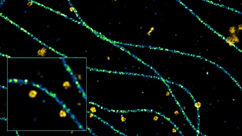
During this webinar, Nicolas will show you how fast and easy it can be to obtain high-quality, reliable, and understandable single-molecule images of your samples with 15 nm resolution in 3D. He will go through the entire imaging workflow—from sample preparation to data analysis—to show you what the single-molecule local microscopy (SMLM) community has made possible, what the common mistakes are, and how Abbelight’s tools and support can make the imaging and analysis journey much easier.

Presenter:

Dr. Nicolas Bourg, Chief Technical Officer and Co-Founder of Abbelight

FAQ

Webinar FAQs | Imaging multiple subcellular structures at the nanometer scale

How long does it take to acquire 3D multicolor nanoscopy images?

The spectral demixing strategy means that acquisition of three-color images typically takes less than one minute for a standard field of view in 3D (about 50 × 50 µm), and a few minutes for larger fields of view (150 × 150 µm).

How difficult is it to prepare nanoscopy structures?

Sample preparation requires only simple modification of the standard preparation procedure, such as changing the concentration of the fixative (e.g., PFA) or increasing the antibody concentration. It only requires fine tuning of your own protocol. The Abbelight team can provide guidance for this if necessary.  

How do you deal with chromatic aberrations in multi-color experiments and with sample drifts?

The spectral demixing strategy allows simultaneous imaging of 2 to 3 far-red channels like 647 nm, 660 nm, and 680 nm. Since the 3 colors are very close in wavelength, there is no chromatic aberrations with this technique.

Sample drifts are always calculated and applied on the image in real time during the acquisition, and they can be refined with different published algorithms and methods in post-acquisition processing.

What is the importance of nanoscopy in diagnosing diseases such as Parkinson’s, Alzheimer’s, and viral infection?

The power of SMLM nanoscopy relies on the fact that the output of an acquisition is not only an image, but a text file with the X,Y, and Z position of every molecule detected during the acquisition. This enables:

  • Quantification of the targeted molecules (up to 3 simultaneously), for instance to follow the evolution of clusters of molecules under drug therapy
  • Nanometric 3D spatial information for structural analysis or colocalization of proteins
  • Tracking of individual molecules in time for dynamic analysis

This potential is currently used to help diagnose diseases, develop novel therapies, diagnose viral or bacterial infection, and much more.

Imaging Multiple Subcellular Structures at the Nanometer Scale in 3D

During this webinar, Nicolas will show you how fast and easy it can be to obtain high-quality, reliable, and understandable single-molecule images of your samples with 15 nm resolution in 3D. He will go through the entire imaging workflow—from sample preparation to data analysis—to show you what the single-molecule local microscopy (SMLM) community has made possible, what the common mistakes are, and how Abbelight’s tools and support can make the imaging and analysis journey much easier.

FAQ

Webinar FAQs | Imaging multiple subcellular structures at the nanometer scale

How long does it take to acquire 3D multicolor nanoscopy images?

The spectral demixing strategy means that acquisition of three-color images typically takes less than one minute for a standard field of view in 3D (about 50 × 50 µm), and a few minutes for larger fields of view (150 × 150 µm).

How difficult is it to prepare nanoscopy structures?

Sample preparation requires only simple modification of the standard preparation procedure, such as changing the concentration of the fixative (e.g., PFA) or increasing the antibody concentration. It only requires fine tuning of your own protocol. The Abbelight team can provide guidance for this if necessary.  

How do you deal with chromatic aberrations in multi-color experiments and with sample drifts?

The spectral demixing strategy allows simultaneous imaging of 2 to 3 far-red channels like 647 nm, 660 nm, and 680 nm. Since the 3 colors are very close in wavelength, there is no chromatic aberrations with this technique.

Sample drifts are always calculated and applied on the image in real time during the acquisition, and they can be refined with different published algorithms and methods in post-acquisition processing.

What is the importance of nanoscopy in diagnosing diseases such as Parkinson’s, Alzheimer’s, and viral infection?

The power of SMLM nanoscopy relies on the fact that the output of an acquisition is not only an image, but a text file with the X,Y, and Z position of every molecule detected during the acquisition. This enables:

  • Quantification of the targeted molecules (up to 3 simultaneously), for instance to follow the evolution of clusters of molecules under drug therapy
  • Nanometric 3D spatial information for structural analysis or colocalization of proteins
  • Tracking of individual molecules in time for dynamic analysis

This potential is currently used to help diagnose diseases, develop novel therapies, diagnose viral or bacterial infection, and much more.

Experts
Dr. Nicolas Bourg
Chief Technical Officer and Co-Founder of Abbelight

Hello. I’m Dr. Nicolas Bourg, chief technical officer (CTO) and co-founder of Abbelight and I’m your designated expert for single molecule localization microscopy (SMLM) – as known as nanoscopy – which includes techniques like PALM, dSTORM, SPT-PALM and DNA-PAINT.

I’m an optronic engineer by training, and I did a Ph.D. in biophotonics at the Paris-Saclay University, working on a unique 3D nanoscopy technique with unprecedented resolution. With my research team, we decided to share all the knowledge I acquired during my PhD and we created Abbelight: a startup that aims to make nanoscopy even more powerful and fully accessible to any biologist without advanced microscopy training. My job is to answer all your questions about nanoscopy, so please get in touch.

Imaging Multiple Subcellular Structures at the Nanometer Scale in 3DDec 13 2024
Sorry, this page is not available in your country
Ask the Expert Sign-up

By clicking subscribe you are agreeing to our privacy policy which can be found here.

Sorry, this page is not
available in your country.

Sorry, this page is not available in your country