IXplore Live
 | Precise Live Cell ImagingDesigned for precise live cell imaging, the IXplore Live microscope system helps reduce photobleaching and enhance cell viability for physiological experiments. |
|---|
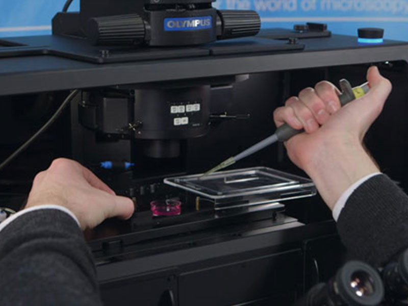 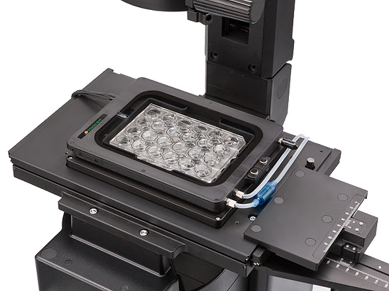  | Meeting the Needs of Live SamplesLive cells require careful maintenance of their environment to grow and thrive, and we offer a variety of microscope-based incubation systems to meet evolving research needs. Box-type incubation systems* enable time-lapse observations over a period of several days by enclosing a portion of the microscope within the incubator. Shorter time-lapse experiments can be completed with stage-top microscope CO2 incubation systems* that can be fitted to the stage and easily removed when not in use. Both systems are precisely controlled to maintain a constant environment surrounding the dish or well plates, controlling temperature, humidity, and CO2 concentration. This maintains cell activity to significantly improve the reliability of time-lapse observation and provide better data. *Third-party products. |
|---|
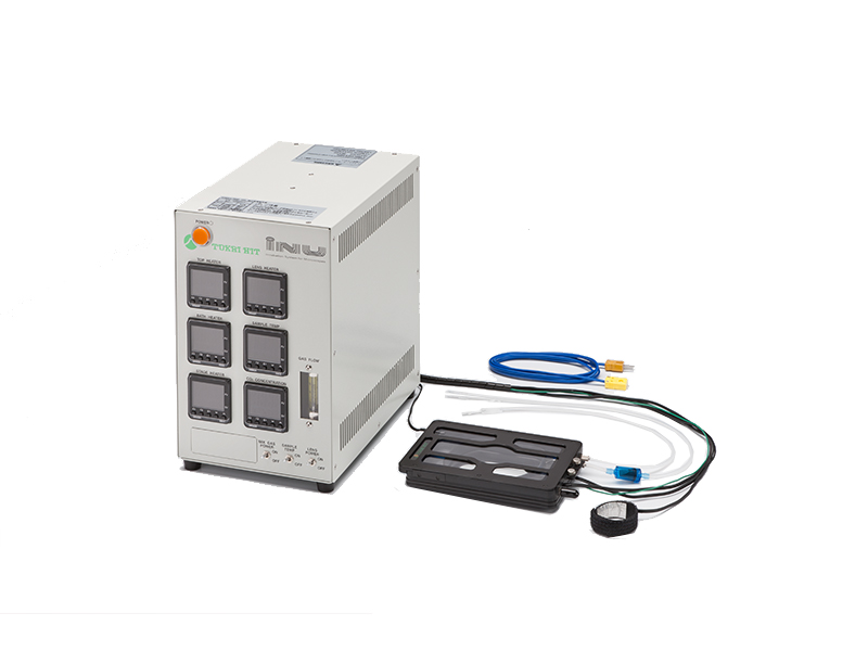
Constant Environmental ControlIn this SelectScience interview, Jutta Bulkescher, Microscopy Specialist at the Center for Protein Research/Danish Stem Cell Center, University of Copenhagen, describes the wide range of research conducted at her facility and explains how the Olympus cellVivo incubation system enables her to reliably perform stem cell analysis, while maintaining cells under strict physiological conditions. |
Hardware StabilityThe frame architecture and focus drive design of the IXplore system offer enhanced rigidity that reduces the impact of vibration and temperature on the microscope. This helps maintain the desired focus position on the Z-axis to facilitate reliable time-lapse imaging. When using the IXplore Live system combined with the TruFocus system, you can capture high-precision time-lapse images that are aligned and in focus. |
|---|
Live Cell ImagingOlympus silicone oil immersion objectives provide clearer images of living specimens during time-lapse experiments. The refractive index of silicone oil (ne≈1.40) is close to that of living tissue (ne≈1.38), so these objectives help reduce the spherical aberration caused by refractive index mismatch, enabling high-resolution observation deep inside living tissue. Silicone oil does not dry out or harden, so there is no need to add more oil, making it ideal for extended time-lapse observations. | 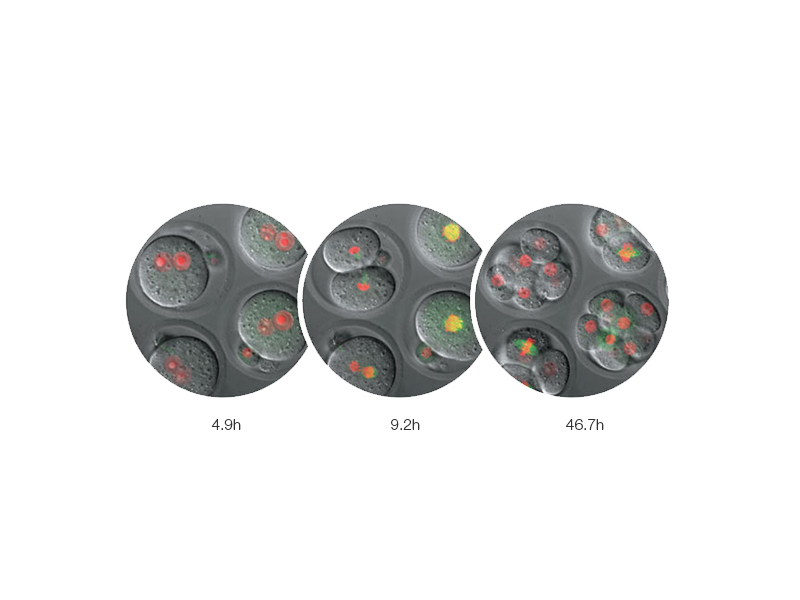 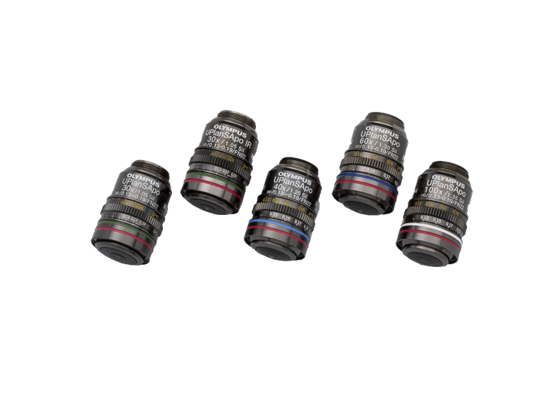 |
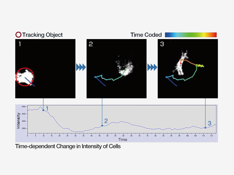 | Monitor Cell Migration and GrowthAnalyze the movement and division of live cells in time-lapse or z-stack image sets with cellSens Object Tracking and Count and Measure solutions. Use the Confluency Checker tools to measure confluency on phase contrast images in addition to fluorescence. |
|---|
Rapid DeconvolutionOlympus cellSens Dimension software includes live 2D deblurring for preview and acquisition to enable better focusing on thick specimens. More advanced TruSight deconvolution is available to reassign out-of-focus light through the CI deconvolution solution. TruSight uses a constrained iterative deconvolution algorithm to produce improved resolution, contrast, and dynamic range with high speed through GPU processing. To improve experiment efficiency, deconvolution processing can be defined as a macro function in the Graphical Experimental Manager (GEM). |  |
|---|
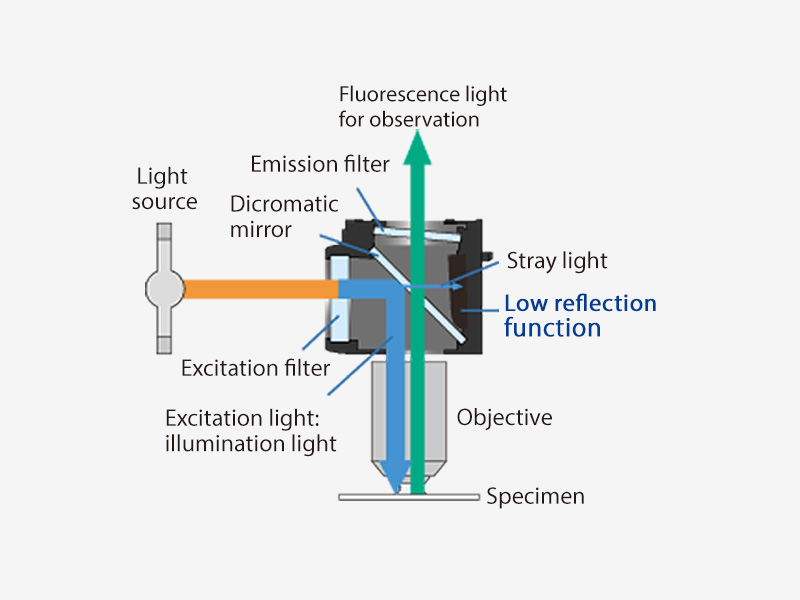 .jpg?rev=8791) | Large Field of ViewThe large field of view Olympus optics, including mirror units and fly-eye lens systems, provide uniformly illuminated fluorescence images and enable the use of sCMOS cameras with large sensors. |
|---|
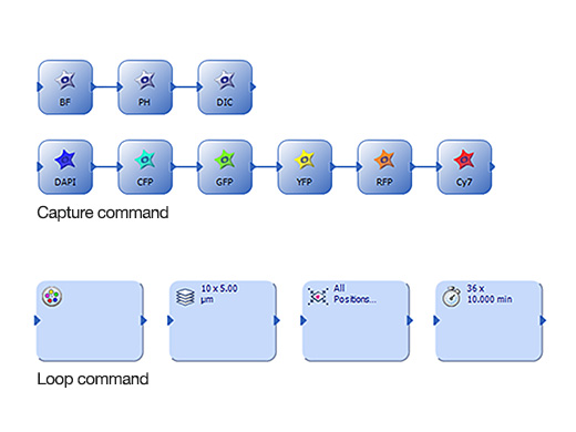
Ease of UseThe Graphical Experimental Manager (GEM) of cellSens Dimension software offers fully automated multidimensional observation (X, Y, Z, T, wavelength, and positions) and eases experiment setup. To increase efficiency, you can define macro functions, such as executing deconvolution processing, in the GEM. |
|
|---|
ReferencesS. Wakayama, et al. Chemical labelling for visualizing native AMPA receptors in live neurons. Nature Communications (April 7, 2017). S. N. Cullati, et al. A bifurcated signaling cascade of NIMA-related kinases controls distinct kinesins in anaphase. The Journal of Cell Biology (June 19, 2017). L. Gheghiani, et al. PLK1 activation in late G2 sets up commitment to mitosis. Cell Reports (June 6, 2017). D. Nakane and T. Nishizaka, et al. Asymmetric distribution of type IV pili triggered by directional light in unicellular cyanobacteria. PNAS (June 5, 2017). T. A. Redchuk, et al. Near-infrared optogenetic pair for protein regulation and spectral multiplexing. Nature Chemical Biology (March 27, 2017). S. Barzilai, et al. Leukocytes breach endothelial barriers by insertion of nuclear lobes and disassembly of endothelial actin filaments. Cell Reports (January 17, 2017). J. Humphries, et al. Species-independent attraction to biofilms through electrical signaling. Cell (January 12, 2017). A. Prindle, et al. Ion channels enable electrical communication in bacterial communities. Nature (October 21, 2015). K. G. Harris, et al. RIP3 regulates autophagy and promotes coxsackievirus B3 infection of intestinal epithelial cells. Cell Host & Microbe (August 13, 2015). |
Need assistance? |
Sorry, this page is not
available in your country.