Not Available in Your Country
Sorry, this page is not
available in your country.
Descripción
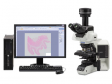
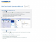
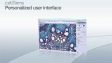
 | Operación intuitiva y proceso de trabajo sin obstáculosLa plataforma cellSens de Olympus proporciona un completo control de la pantalla y de la ubicación de los íconos, barras de herramientas y comandos. Esto permite que el software se estructure y adapte según sus necesidades de investigación en constante evolución. |
|---|
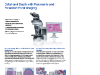
Paquetes cellSens |
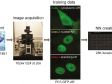
cellSens EntrycellSens Entry es el trampolín ideal para los investigadores que quieran adentrarse en la adquisición y documentación de imágenes digitales, ya que proporciona todas las herramientas necesarias para la adquisición sencilla de imágenes. |
*cellSens Entry no está disponible en algunas regiones. |
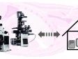
cellSens StandardLa versión del software Olympus cellSens Standard está modelada en función del paquete cellSens Entry, llevando la adquisición más allá de una sola imagen, con procesos avanzados de captura de imágenes (como intervalos) y control de componentes de microscopios motorizados y codificados. |
|
cellSens DimensionEl miembro más versátil de la familia cellSens de Olympus es el cellSens Dimension, con una adquisición de imágenes completamente automática, potentes herramientas de análisis y mucho más. |
|
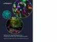
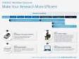
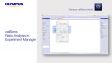
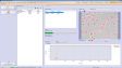
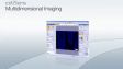
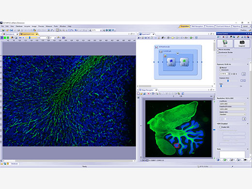 | Adquisición experimental 5DPermite adquirir imágenes en cinco dimensiones empleando herramientas, como el Graphic Experiment Manager (GEM) y el Well Navigator para ayudar a visualizar la adquisición de sus datos de forma sencilla. |
|---|
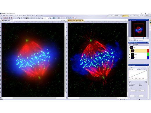
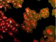
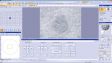
Procesamiento e intercambio de imágenesRevele datos fiables de sus imágenes gracias a la técnica de procesamiento de imagen TruSight deconvolution y otras más. Comparta con facilidad sus resultados con otras personas al usar el parámetro Conference Mode, o arrastre y coloque sus datos en informes preconfigurados. |
|
|---|
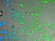
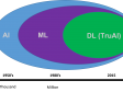
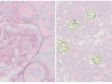
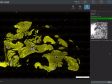
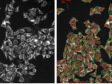
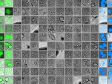
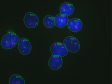
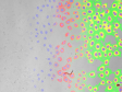
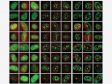
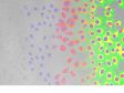
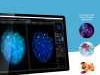
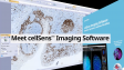
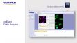
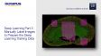
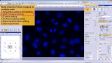
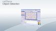
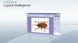
| Potentes herramientas de análisisTrabaje de forma dinámica con sus imágenes para adquirir todos los datos posibles que le ayudarán a obtener resultados experimentales fiables. La tecnología de aprendizaje profundo del software (TruAI) ofrece un análisis de segmentación mejorado. Use la función Macro Manager para automatizar los flujos completos de trabajo a lo largo de todo el análisis y almacenamiento de sus imágenes. |
|---|

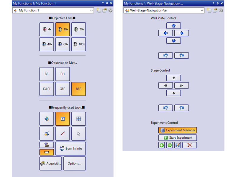
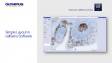
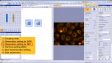
Interfaz del usuario personalizableSeleccione un diseño de pantalla recomendado para la adquisición y análisis de sus imágenes o cree su propio diseño usando el conjunto de herramientas My Functions. |
|
|---|
¿Necesita ayuda? |
El software cellSens no está diseñado para fines de diagnóstico clínico. |
Especificaciones
Funciones y soluciones opcionales del software cellSens |
| Dimension | Standard | Entry | |||
|---|---|---|---|---|---|
| Diseño de pantalla | Personalización de la experiencia del usuario | ✓ | ✓ | ✓ | |
| Visualización | Superposición de varias imágenes | ✓ | ✓ | - | |
| Grupos de documentos para comparar imágenes lado a lado | ✓ | ✓ | ✓ | ||
| Reproducción de videos | ✓ | ✓ | ✓ | ||
| Vista en mosaico (varias imágenes de un mismo conjunto de datos mostradas lado a lado) | ✓ | ✓ | ✓ | ||
| Vista transversal para visualizar el plano ortogonal de conjuntos de datos 3D o intervalos | ✓ | - | - | ||
| Visor de vóxeles para la representación volumétrica y de isosuperficies de conjuntos de datos 3D y 4D | ✓ | - | - | ||
| Adquisición de imágenes | Adquisición de instantáneas/video | ✓ | ✓ | ✓ | |
| Período de tiempo a intervalos específicos | ✓ | ✓ | - | ||
| Longitud de onda múltiple automatizada | ✓ | ✓ | - | ||
| Apilamiento en Z | ✓ | - | - | ||
| Multidimensional (XYZT y longitud de onda) | ✓ | - | - | ||
| Administrador Gráfico de Experimentos (GEM: Graphical Experiment Manager) | ✓ | - | - | ||
| Procesamiento manual de imágenes panorámicas (MIA intantánea y MIA manual) | ✓ | Proceso manual | Proceso manual | ||
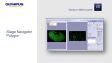
| Inspección multiposición y navegador de platina | Multiposición | Multiposición | - | ||
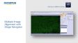
| Creación automática de imágenes panorámicas (MIA automático; requiere platina motorizada) | Multiposición | Multiposición | - | ||
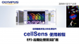
| Creación instantánea de imagen EFI (eje Z manual o motorizado) | ✓ | Proceso manual | Proceso manual | ||
| Procesamiento simultáneo de imágenes multicolor (requiere dos cámaras** idénticas o un divisor de imágenes) | ✓ | - | - | ||
| Corrección de desenfoque en vivo | ✓ | - | - | ||
| Creación de imágenes de amplio rango dinámico (HDRI) | ✓ | - | - | ||
| Collar de corrección automático (ACC) | ✓ | ✓ | - | ||
| Adquisición de placas con múltiples pocillos | Navegador de placa de pocillos y Multiposición | - | - | ||
| Procesamiento de imágenes | Procesamiento por filtros/combinación/geometría | ✓ | ✓ | - | |
| Separación espectral en fluorescencia | ✓ | - | - | ||
| Separación espectral en campo claro | Recuento y medición | - | - | ||
| Corrección de desenfoque (sin proximidad/proximidad más cercana, filtro Wiener) | ✓ | - | - | ||
| Quimógrafo | ✓ | - | - | ||
| Deconvolución 2D | ✓ | - | - | ||
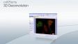
| Deconvolución 3D (deconvolución iterativa limitada con procesamiento en GPU) | Deconvolución iterativa limitada | - | - | ||
| Análisis de imágenes | Análisis de fase | ✓ | - | - | |
| Medición y clasificación de objetos | Recuento y medición | Recuento y medición | - | ||
| Mediciones 2D interactivas | ✓ | ✓ | ✓* | ||
| Gráfico de intensidad en función del tiempo/z | ✓ | - | - | ||
| Colocalización | ✓ | - | - | ||
| Conteo de objetos (manual) | ✓ | ✓ | - | ||
| Seguimiento de objetos | Seguimiento y Recuento y medición | - | - | ||
| Ratio y cinética en línea | Ratio/FRET | - | - | ||
| Análisis del ratio (fuera de línea) | ✓ | - | - | ||
| Análisis FRET | Ratio/FRET o Análisis de ciencias de la vida | - | - | ||
| Análisis FRAP | Fotomanipulación o Análisis de ciencias de la vida | - | - | ||
| Recuento celular y medición de confluencia | ✓ | Verificador de confluencia | - | ||
| Aprendizaje profundo (TruAI) | Formación de redes neurales | Aprendizaje profundo (TruAI) | Aprendizaje profundo (TruAI) | - | |
| Inferencia usando redes neurales formadas (en línea/fuera de línea) | Aprendizaje profundo (TruAI) o Recuento y medición | Aprendizaje profundo (TruAI) o Recuento y medición | - | ||
| Documentación y colaboración | Composición automática de informes en MS Word | ✓ | - | - | |
| Base de datos para la administración de imágenes y datos de microscopía | Base de datos Core | Base de datos Core | - | ||
| Abrir base de datos y cargar registros/documentos en base de datos | Base de datos Client | Base de datos Client | Base de datos Client | ||
| Comunicación remota | Visualización remota de imágenes en vivo | NetCam | NetCam | - | |
| * Solamente ángulo de tres puntos, ángulo de cuatro puntos, línea arbitraria, polígono cerrado, polilínea y línea perpendicular. La opción de mediciones 2D interactivas es requerida para agregar otras herramientas de medición y habilitar la exportación a hojas de cálculo Excel.
** Cámaras soportadas: iXon Ultra 897; Zyla 5.5 (USB 3.0); Zyla 4.2 (USB 3.0/CamLink); Neo, iXon Ultra 888, ImagEM X2; ORCA-Flash 4.0 (V2/V3); Prime 95B; Prime BSI; Prime BSI Express; Sona4.2B-11; ORCA Fusion; ORCS-Fusion BT; ORCA-QUEST. |
| Dimension | Standard | |||
|---|---|---|---|---|
| Diseño de pantalla | Personalización de la experiencia del usuario | ✓ | ✓ | |
| Control del microscopio | Control del microscopio | ✓ | ✓ | |
| Visualización | Vista transversal para visualizar el plano ortogonal de conjuntos de datos 3D o intervalos | ✓ | - | |
| Visor de vóxeles para la representación volumétrica y de isosuperficies de conjuntos de datos 3D y 4D | ✓ | - | ||
| Adquisición de imágenes | Longitud de onda múltiple automatizada | ✓ | ✓ | |
| Apilamiento en Z | ✓ | - | ||
| Multidimensional (XYZT y longitud de onda) | ✓ | - | ||
| Creación instantánea de imagen EFI (eje Z manual o motorizado) | ✓ | Proceso manual | ||
| Procesamiento automático de imágenes panorámicas (MIA automática; requiere platina motorizada) | Multiposición | Multiposición | ||
| Procesamiento manual de imágenes panorámicas (MIA intantánea y MIA manual) | ✓ | Proceso manual | ||
| Procesamiento de imágenes simultáneo y multicromático (requiere dos cámaras idénticas o un divisor de imágenes)*1 | ✓ | - | ||
| Corrección de desenfoque en vivo | ✓ | - | ||
| Procesamiento de alto rango dinámico (HDR) | ✓ | - | ||
| Collar de corrección automático (ACC) | ✓ | ✓ | ||
| Adquisición de placas con múltiples pocillos | Navegador de placa de pocillos y Multiposición | - | ||
| Procesamiento de imágenes | Alineación de múltiples imágenes (MIA) | ✓ | ✓ | |
| Procesamiento por filtros/combinación/geometría | ✓ | ✓ | ||
| Filtro morfológico | Recuento y medición | Recuento y medición | ||
| Separación espectral en fluorescencia | ✓ | - | ||
| Separación espectral en campo claro | Recuento y medición | - | ||
| Quimógrafo | ✓ | - | ||
| Deconvolución 2D | ✓ | - | ||
| Deconvolución 3D (deconvolución iterativa restringida) | Deconvolución iterativa limitada | - | ||
| Análisis de imágenes | Mediciones 2D interactivas | ✓ | ✓ | |
| Conteo de objetos (manual) | ✓ | ✓ | ||
| Colocalización | ✓ | - | ||
| Medición y clasificación de objetos | Recuento y medición | Recuento y medición | ||
| Seguimiento de objetos | Seguimiento y Recuento y medición | - | ||
| Ratio y cinética en línea | Ratio/FRET | - | ||
| Análisis de ratio (fuera de línea) | ✓ | - | ||
| Análisis FRET | Ratio/FRET o Análisis de ciencias de la vida | - | ||
| Análisis FRAP | Análisis para las ciencias de la vida | - | ||
| Recuento celular y medición de confluencia | ✓ | Verificador de confluencia | ||
| Aprendizaje profundo (TruAI) | Formación de redes neurales | Aprendizaje profundo (TruAI) | Aprendizaje profundo (TruAI) | |
| Inferencia usando redes neurales formadas (en línea/fuera de línea) | Aprendizaje profundo (TruAI) o Recuento y medición | Aprendizaje profundo (TruAI) o Recuento y medición | ||
| Informes | Función de generación de informes (requiere Microsoft Word) | ✓ | - | |
| Documentación y colaboración | Base de datos para la administración de imágenes y datos de microscopía | Base de datos Core | Base de datos Core | |
| Abrir base de datos y cargar registros/documentos en base de datos | Base de datos Client | Base de datos Client | ||
* Cámaras soportadas: iXon Ultra 897; Zyla 5.5 (USB 3.0); Zyla 4.2 (USB 3.0/CamLink); Neo, iXon Ultra 888, ImagEM X2; ORCA-Flash 4.0 (V3); Prime 95B; Prime BSI; Prime BSI Express; Sona4.2B-11; ORCA-Fusion; ORCA-Fusion BT; ORCA-QUEST. |
Soluciones cellSens■ Incluida □ Opcional |
| Dimension | Standard | Entry | |||
|---|---|---|---|---|---|
| Proceso manual | [Manual Process] Cree fácilmente imágenes de compuestos de alta resolución (en MIA instantánea) al mover simplemente la platina manual. También es posible adquirir una imagen focal extendida (EFI) sobre la superficie completa al desviar manualmente la dirección Z. | ■ | □ | □ | |
| Dispositivo codificado | [Encoded devices] Dispositivos codificados (objetivos, intensidad de luz, etc.) que facilitan la recuperación de configuraciones. | ■ | ■ | □ | |
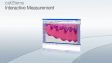
| Medición interactiva | [Interactive Measurement] Dibuje una polilínea, un rectángulo o un círculo sobre su imagen para obtener datos de medición exportables. Los resultados de medición pueden ser exportados a una hoja Excel. | ■ | ■ | □ | |
| Base de datos Client | [Database Client] Da acceso a la base de datos creada con la opción Database Core. | □ | □ | □ | |
| Base de datos Core | [Database Core] Haga que la administración de los datos y su navegación a través de ellos sea más eficiente creando una base de datos a partir de la cual pueda buscar y clasificar fácilmente las imágenes adquiridas en función de los datos, como las condiciones de captura y la fecha de adquisición. | □ | □ | ||
| Verificador de confluencia | [Confluency Checker] Permite determinar la confluencia de células vivas sin tinción en recipientes de cultivo mediante mediciones cuantitativas para obtener datos fiables. | ■ | □ | ||
| Multiposición | [Multiposition] Las imágenes de múltiples áreas y aquellas unidas en mosaico pueden adquirirse usando la platina motorizada. Al ser combinadas con el enfoque motorizado en Z, es posible crear un mapa de enfoque a partir de múltiples puntos enfocados; asimismo, es posible obtener imágenes unidas con poca desviación de enfoque eliminando la inclinación y la distorsión de la muestra. | □ | □ | ||
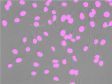
| Recuento y medición | [Count & Measure] Defina la morfología de un objeto, y el software identificará todos losobjetos similares y presentará los resultados del análisis de segmentación en un gráfico. | □ | □ | ||
| NetCam | Facilita la transferencia de imágenes en vivo y almacenadas a través de una red destinadas a la docencia, enseñanza, tutoría o supervisión. | □ | □ | ||
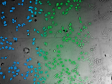
| Aprendizaje profundo | [Deep Learning] Eficiente análisis de segmentación estimulado por el aprendizaje profundo que permite la detección de objetivos difíciles, como la detección de núcleos sin marcado. | □ | □ | ||
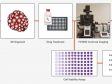
| Navegador de placa de pocillos*1 | [Well Plate Navigator] Configure fácilmente la captura para cada pocillo. La posición y el nombre del pocillo pueden ser etiquetados a imágenes, lo que facilita la gestión de losdatos y hace que la inspección de la placa de pocillos sea más eficiente. | □ | |||
| Deconvolución iterativa limitada | [CI Deconvolution] Acceda al GPU basado en la deconvolución así como a los populares y personalizados algoritmos de deconvolución TruSight para mejorar la nitidez, el contraste y el rango dinámico de las imágenes reconstruidas. | □ | |||
| Ratio/FRET | Obtenga medidas de cocientes a partir de sus imágenes, a medida que van siendo capturadas. | □ | |||
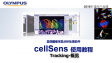
| Seguimiento*2 | [Tracking] Mida y analice la luminancia y la velocidad de las células individuales que se mueven y se dividen a través del tiempo. | □ | |||
| Análisis para las ciencias de la vida | [Life Science Analysis] Es posible ejecutar análisis de recuperación de fluorescencia después del fotoblaqueo (FRAP) y de transmisión de energía de resonancia de fluorescencia (FRET) en la imagen adquirida. | □ | |||
| Fotomanipulación | [Photo Manipulation] Habilita el control del módulo cellFRAP y el análisis FRAP. | □ | |||
| Control láser | Permite a NI USB-6343 BNC controlar dispositivos externos. | □ | |||
| Collar de corrección automático (ACC) | Permite operar automáticamente el collar de corrección. | □ | □ | ||
| *1 Requiere la opción Multiposición [Multiposition]
*2 Requiere la opción de Recuento y medición [Count & Measure] |
| Dimension | Standard | |||
|---|---|---|---|---|
| Proceso manual | [Manual Process] Cree fácilmente imágenes de compuestos de alta resolución (en MIA instantánea) al mover simplemente la platina manual. También es posible adquirir una imagen focal extendida (EFI) sobre la superficie completa al desviar manualmente la dirección Z. | ■ | □ | |
| Dispositivo codificado | [Encoded devices] Dispositivos codificados (objetivos, intensidad de luz, etc.) que facilitan la recuperación de configuraciones. | ■ | ■ | |
| Medición interactiva | [Interactive Measurement] Dibuje una polilínea, un rectángulo o un círculo sobre su imagen para obtener datos de medición exportables. Los resultados de medición pueden ser exportados a una hoja Excel. | ■ | ■ | |
| Base de datos Client | [Database Client] Da acceso a la base de datos creada con la opción Database Core. | □ | □ | |
| Base de datos Core | [Database Core] Haga que la administración de los datos y su navegación a través de ellos sea más eficiente creando una base de datos a partir de la cual pueda buscar y clasificar fácilmente las imágenes adquiridas en función de los datos, como las condiciones de captura y la fecha de adquisición. | □ | □ | |
| Verificador de confluencia | [Confluency Checker] Permite determinar la confluencia de células vivas sin tinción en recipientes de cultivo mediante mediciones cuantitativas para obtener datos fiables. | ■ | □ | |
| Multiposición | [Multiposition] Las imágenes de múltiples áreas y aquellas unidas en mosaico pueden adquirirse usando la platina motorizada. Al ser combinadas con el enfoque motorizado en Z, es posible crear un mapa de enfoque a partir de múltiples puntos enfocados; asimismo, es posible obtener imágenes unidas con poca desviación de enfoque eliminando la inclinación y la distorsión de la muestra. | □ | □ | |
| Recuento y medición | [Count & Measure] Defina la morfología de un objeto, y el software identificará todos losobjetos similares y presentará los resultados del análisis de segmentación en un gráfico. | □ | □ | |
| NetCam | Facilita la transferencia de imágenes en vivo y almacenadas a través de una red destinadas a la docencia, enseñanza, tutoría o supervisión. | □ | □ | |
| Aprendizaje profundo | [Deep Learning] Eficiente análisis de segmentación estimulado por el aprendizaje profundo que permite la detección de objetivos difíciles, como la detección de núcleos sin marcado. | □ | □ | |
| Navegador de placa de pocillos*1 | [Well Plate Navigator] Configure fácilmente la captura para cada pocillo. La posición y el nombre del pocillo pueden ser etiquetados a imágenes, lo que facilita la gestión de losdatos y hace que la inspección de la placa de pocillos sea más eficiente. | □ | ||
| Deconvolución iterativa limitada | [CI Deconvolution] Acceda al GPU basado en la deconvolución así como a los populares y personalizados algoritmos de deconvolución TruSight para mejorar la nitidez, el contraste y el rango dinámico de las imágenes reconstruidas. | □ | ||
| Ratio/FRET | Obtenga medidas de cocientes a partir de sus imágenes, a medida que van siendo capturadas. | □ | ||
| Seguimiento*2 | [Tracking] Mida y analice la luminancia y la velocidad de las células individuales que se mueven y se dividen a través del tiempo. | □ | ||
| Análisis para las ciencias de la vida | [Life Science Analysis] Es posible ejecutar análisis de recuperación de fluorescencia después del fotoblaqueo (FRAP) y de transmisión de energía de resonancia de fluorescencia (FRET) en la imagen adquirida. | □ | ||
| Fotomanipulación | [Photo Manipulation] Habilita el control del módulo cellFRAP y el análisis FRAP. | □ | ||
| Control láser | Permite a NI USB-6343 BNC controlar dispositivos externos. | □ | ||
| Collar de corrección automático (ACC) | Permite operar automáticamente el collar de corrección. | □ | □ | |
| *1 Requiere la opción Multiposición [Multiposition]
*2 Requiere la opción de Recuento y medición [Count & Measure] |
Productos con funcionalidad confirmada |
| Dimension | Standard | Entry | |||
|---|---|---|---|---|---|
| Olympus | Cámaras | DP23; DP23M; DP28; DP74; DP75; DP80; XM10; UC90; LC20; LC30; LC35; SC50; SC180 | ✓ | ✓ | ✓ |
| Microscopio | BX43; BX53; BX63; BX61; BX61WI; IX83; IX85; IX73; IX81; SZX16A | ✓ | ✓ | - | |
| IX81-ZDC; IX81-ZDC2 | ✓ | - | - | ||
| Periféricos | BX-DSU; IX3-DSU; IX3-ZDC; IX3-ZDC2; IX2-DSU; U-CBF; cellTIRF (multilínea, línea única); convertidor USB-ODB; controlador instantáneo (U-RTCE); IX5-ZDC | ✓ | - | - | |
| Fuente de luz | U-LGPS | ✓ | ✓ | - | |
| Hamamatsu | Cámaras | ImagEMX2; ORCA-Flash 4.0 V3; ORCA-Flash 4.0 LT PLUS; ORCA-Flash 4.0 LT3; ORCA-Fusion; ORCA-Fusion BT; ORCA-QUEST | ✓ | - | - |
| ORCA-spark | ✓ | ✓ | - | ||
| Divisor de imágenes | W-View Gemini | ✓ | - | - | |
| Q-Imaging | Cámaras | Retiga 6000 | ✓ | - | - |
| Photometrics | Cámaras | Prime (PCI-Express); Prime 95B; Prime BSI; Prime BSI Express; Moment | ✓ | - | - |
| Divisor de imágenes | Dual View DV2 / QuadView QV2 | ✓ | - | - | |
| Andor | Cámaras | iXon Ultra 897; iXon Ultra 888; iXon Life 888; iXon Life 897; Sona4.2B-11 Zyla4.2/Zyla4.2 PLUS (Camera-link,USB3.0); Zyla5.5 (Camera-link 10tap,USB3.0); ZL41 Cell 4.2 (Camera-link,USB3.0); Neo5.5 | ✓ | - | - |
| Vincent Associates | Obturador | Uniblitz shutter (VCM-D1, VMM-D1, VMM-D3) | ✓ | ✓ | - |
| CoolLED | Fuente de luz | pE-1; pE-2; pE800; pE-4000 | ✓ | - | - |
| pE-300white; pE-300ultra; pE-340fura | ✓ | ✓ | - | ||
| Excelitas | Fuente de luz | X-Cite120LED; X-Cite XYLIS; X-Cite TURBO | ✓ | - | - |
| Lumencor | Fuente de luz | SOLA SEII; SEII 365; Spectra X | ✓ | - | - |
| Sutter | Obturador, rueda de filtros (FW) | Lambda 10-3/10-B | ✓ | - | - |
| Prior | Platina XY motorizada | ProScan III; Optiscan III | Multiposición | - | - |
| Obturador, rueda de filtros (FW), sistema de desplazamiento en Z | ProScan (I, II, III); Optiscan III | ✓ | - | - | |
| Piezo Z (control a través del controlador instantáneo) | NanoScanZ NZ100 | ✓ | - | - | |
| Ludl | Platina XY motorizada | Mac 6000 | Multiposición | - | - |
| Obturador, rueda de filtros (FW), sistema de desplazamiento en Z | Mac 6000 | ✓ | - | - | |
| Märzhäuser | Platina XY motorizada | Platinas Tango; Pilot | Multiposición | - | - |
Controlador de sistema de desplazamiento en Z | Tango | ✓ | - | - | |
| Physik Instrumente | Piezo Z (control a través del controlador instantáneo) | PIFOC P-721 | ✓ | - | - |
| Applied Scientific Instrumentation | Platina XY motorizada | MS-2000 | Multiposición | - | - |
| Controlador de sistema de desplazamiento en Z | MS-2000 | ✓ | - | - | |
| National Instruments | Dispositivo TTL digital | NI USB-6501 | ✓ | - | - |
| NI USB-6343 BNC | Control láser | - | - | ||
| Yokogawa | CSU | CSU-X1, CSU-W1 | ✓ | - | - |
| Para más información con respecto a la compatibilidad del sistema operativo (OS) de Windows, póngase en contacto con el representante local de ventas de Evident. |
| Dimension | Standard | |||
|---|---|---|---|---|
| Olympus | Cámaras | DP23; DP23M; DP28; DP74; DP75; DP80 | ✓ | ✓ |
| Microscopio | BX43; BX53; BX63; BX61; BX61WI; IX83; IX85; IX73; IX81; SZX16A | ✓ | ✓ | |
| IX81-ZDC; IX81-ZDC2 | ✓ | - | ||
| Periféricos | BX-DSU; IX3-DSU; IX3-ZDC; IX3-ZDC2; IX2-DSU; U-CBF; controlador instantáneo (U-RTCE); IX5-ZDC | ✓ | - | |
| Fuente de luz | U-LGPS | ✓ | ✓ | |
| Hamamatsu | Cámaras | ImagEMX2; ORCA-Flash 4.0 V3; ORCA-Flash 4.0 LT PLUS; ORCA-Fuision, ORCA-Fusion BT; ORCA-QUEST | ✓ | - |
| ORCA-spark | ✓ | ✓ | ||
| Divisor de imágenes | W-View Gemini | ✓ | - | |
| Q-Imaging | Cámaras | Retiga 6000 | ✓ | - |
| Photometrics | Cámaras | Prime; Prime 95B; Prime BSI; Prime BSI Express; Moment | ✓ | - |
| Divisor de imágenes | Dual View DV2 / QuadView QV2 | ✓ | - | |
| Andor | Cámaras | iXon Ultra 897; iXon Ultra 888; iXon Life 888; iXon Life 897; Sona4.2B-11 Zyla4.2/Zyla4.2 PLUS (Camera-link,USB3.0); Zyla5.5 (Camera-link 10tap,USB3.0); ZL41 Cell 4.2 (Camera-link,USB3.0); Neo5.5 | ✓ | - |
| Vincent Associates | Obturador | Uniblitz shutter (VCM-D1, VMM-D1, VMM-D3) | ✓ | ✓ |
| Ludl | Platina XY motorizada | Mac 6000 | Multiposición | - |
| Obturador, rueda de filtros (FW), sistema de desplazamiento en Z | Mac 6000 | ✓ | - | |
| Prior | Platina XY motorizada | ProScan III; Optiscan III | Multiposición | - |
| CoolLED | Fuente de luz | pE-1; pE-2; pE800; pE-4000 | ✓ | - |
| pE-300white; pE-300ultra; pE-340fura | ✓ | ✓ | ||
| Excelitas | Fuente de luz | X-Cite120LED; X-Cite XYLIS; X-Cite TURBO | ✓ | - |
| Lumencor | Fuente de luz | SOLA SEII; SEII 365; Spectra X | ✓ | - |
| Sutter | Obturador, rueda de filtros (FW) | Lambda 10-3/10-B | ✓ | - |
| National Instruments | Dispositivo TTL digital | NI USB-6501 | ✓ | - |
| NI USB-6343 BNC | Control láser | - | ||
| Yokogawa | CSU | CSU-W1 | ✓ | - |
| Para más información con respecto a la compatibilidad del sistema operativo (OS) de Windows, póngase en contacto con el representante local de ventas de Evident. |
Formatos de imagen compatibles |
| Lectura y escritura | JPEG; JPEG2000; TIFF; BMP; AVI; PNG; VSI; PSD (Adobe Photoshop); Big TIFF; OIR | ||||
|---|---|---|---|---|---|
| Sólo lectura | GIF; OIF/OIB (formato FLUOVIEW); Cell; STK (MetaMorph); MRC (Medical Research Council) | ||||
Requisitos del sistema |
| Sistema operativo | Microsoft Windows 10 Professional (64 bits) (22H2), Microsoft Windows 11 Pro (64 bits) (23H2) | ||||
|---|---|---|---|---|---|
| Idioma del sistema operativo | Inglés, chino simplificado, japonés, alemán e italiano (Entry y Standard) | ||||
| CPU | Intel Core i5; Intel Core i7; Intel Core i9; Intel Xeon que es recomendado para la adquisición de imágenes a alta velocidad: QuadCore | ||||
| RAM | 8 GB para las aplicaciones generales; se recomienda 16 GB o más para la adquisición de imágenes de alta velocidad [para las cámaras DP23/DP28/DP23M, se recomienda una memoria dual para un procesamiento de imágenes de alto refresco de fotogramas); se recomienda 32 GB o más para el aprendizaje profundo. | ||||
| Disco duro |
5 GB para instalación
Recomendado para la adquisición de imágenes en alta velocidad: unidad de estado sólido (SSD) | ||||
Actualización de la versión del software
Una actualización de la versión de software está disponible para la versión posterior a la especificada en la tarjeta de licencia (se excluyen las versiones anteriores).
|

![cellSens [ver.4.3.1] Hardware Manual](https://static5.olympus-lifescience.com/modules/imageresizer/ff7/926/48f03e1b99/100x75p50x71.jpg)
![cellSens [ver.4.3.1] Database Manual](https://static4.olympus-lifescience.com/modules/imageresizer/e7e/c9a/f7d7ac0695/100x75p50x72.jpg)
![cellSens [ver.4.3.1] User Manual](https://static5.olympus-lifescience.com/modules/imageresizer/725/338/b98fb8d335/100x75p50x71.jpg)
![cellSens [ver.4.3.1] Installation Manual](https://static4.olympus-lifescience.com/modules/imageresizer/03c/ef4/1f631b053d/100x75p50x71.jpg)
![cellSens [ver.4.3] User Manual](https://static3.olympus-lifescience.com/modules/imageresizer/80a/c74/e7eac6d841/100x75p50x71.png)
![cellSens [ver.4.3] Database Manual](https://static1.olympus-lifescience.com/modules/imageresizer/a50/21c/a66418e8b6/100x75p50x72.png)
![cellSens [ver.4.3] Installation Manual](https://static5.olympus-lifescience.com/modules/imageresizer/855/a64/34311544eb/100x75p50x74.png)
![cellSens [ver.4.3] Hardware Manual](https://static2.olympus-lifescience.com/modules/imageresizer/ee3/c64/b4b75fddd2/100x75p50x74.png)
![cellSens [ver.4.2.1] User Manual](https://static5.olympus-lifescience.com/modules/imageresizer/64e/8c7/4132b5b5e5/112x84p74x50.jpg)
![cellSens [ver.4.2.1] Installation Manual](https://static2.olympus-lifescience.com/modules/imageresizer/99b/1ba/44507be555/112x84p63x50.jpg)
![cellSens [ver.4.2.1] Hardware Manual](https://static3.olympus-lifescience.com/modules/imageresizer/241/054/8e949a7a55/112x84p69x50.jpg)
![cellSens [ver.4.2.1] Database Manual](https://static5.olympus-lifescience.com/modules/imageresizer/254/161/ab9aae4bef/112x84p67x50.jpg)
![cellSens [ver.4.2] Database Manual](https://static4.olympus-lifescience.com/modules/imageresizer/051/014/b73c664ebb/112x84p63x50.jpg)
![cellSens [ver.4.2] Installation Manual](https://static5.olympus-lifescience.com/modules/imageresizer/1e2/f4e/59fde3cca6/112x84p69x50.jpg)
![cellSens [ver.4.2] Hardware Manual](https://static3.olympus-lifescience.com/modules/imageresizer/ed0/00b/da9613f82e/112x84p67x50.jpg)
![cellSens [ver.4.2] User Manual](https://static3.olympus-lifescience.com/modules/imageresizer/31c/7b4/c7022777f0/112x84p71x50.jpg)
![cellSens [ver.4.1] Installation Manual](https://static4.olympus-lifescience.com/modules/imageresizer/d67/038/32f7f54871/112x84p61x50.jpg)
![cellSens [ver.4.1] Hardware Manual](https://static3.olympus-lifescience.com/modules/imageresizer/626/e28/7713ca10bb/112x84p68x50.jpg)

![cellSens [ver.4.1] Database Manual](https://static2.olympus-lifescience.com/modules/imageresizer/a4d/346/5e7903657a/112x84p63x50.jpg)
![cellSens [ver.4.1] User Manual](https://static5.olympus-lifescience.com/modules/imageresizer/15a/70a/01d83866c2/112x84p61x50.jpg)


Descargas
Installers and Version Checker |
cellSens V4.3.1 64-Bit Installer | cellSens V4.3 64bit Installer | cellSens V4.2.1 64bit Installer |
cellSens V4.2 64bit Installer | cellSens V4.1.1 64bit Installer | cellSens V3.2 64bit Installer |
cellSens V2.3 32bit InstallercellSens V2.3 64bit Installer | cellSens V1.18 32bit InstallercellSens V1.18 64bit Installer | cellSens V1.16 32bit InstallercellSens V1.16 64bit Installer |
VERSION 1.7 OR LATER | Upgrading to a Windows 10 PC | |
Notas de version
Version 4.3.1New Hardware Support
New Functions and Improvements
Version 4.3New function/improvement
Version 4.2.1New hardware support
New function/improvement
Version 4.2New hardware support
New function/improvement
Version 4.1.1New hardware support
Minor bug improvements
Version 4.1New hardware support
New function/improvement
Version 3.2New hardware support
New function/improvement
Version 2.3New hardware support
New function/improvement
Version 2.2New hardware support
New function/improvement
Version 2.1New hardware support
Product portfolio changes
New function/improvement
|