Introduction to Laser Scanning Microscopes
Laser scanning microscopes use laser illumination to generate high-resolution, high-contrast 3D imagery of samples by scanning them point by point. Two common types of laser scanning microscopes include confocal laser scanning microscopes and multiphoton laser scanning microscopes. While both use lasers to excite the sample, there are some key differences between them. Here we will explain how both confocal laser scanning microscopes and multiphoton laser scanning microscopes work, their uses, and the types of images they can create.
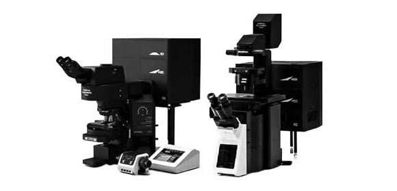
Confocal Laser scanning microscopes |
Multiphoton laser scanning microscopes |
Principles of Confocal Laser Scanning Microscopes
Confocal laser scanning microscopes work by using a short wavelength continuous wave (CW) laser to create high-intensity excitation light to illuminate the sample. This laser is reflected off a dichroic mirror and then two more mirrors that, in turn, scan the laser across a dyed sample. When excited by the laser, the dye in the sample fluoresces and emits a long wavelength light that is descanned by the mirrors previously used to scan the excitation light. The emitted light then passes through the dichroic mirror, where it is focused onto a pinhole and collected and measured by the detector. The detector is connected to a computer that builds a digital 3D image point by point. Since the pinhole blocks out any out-of-focus fluorescence, the image created is highly detailed. The principle of confocal laser scanning microscopy is that both the laser and detector are focused on the same point of the sample, and this point shifts to ultimately build a complete image of the sample. |
Principles of Multiphoton Laser Scanning Microscopes
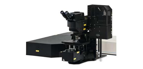
Multiphoton laser scanning microscopes work in a very similar manner to confocal laser scanning microscopes. However, while confocal laser scanning microscopes use a short visible wavelength laser to excite the sample, multiphoton laser scanning microscopy uses a femtosecond infrared (IR) pulse laser emitted at wavelengths two times longer than what is required for single-photon excitation. When the dye in the sample absorbs two photons simultaneously, it can emit fluorescence. This two-photon absorption phenomenon only happens at the focus position where the photon density is highly concentrated, and only the dye at the focal point can be excited and emits fluorescence. Hence, a multiphoton laser scanning microscope system does not require a pinhole aperture to block out-of-focus fluorescence for its nondescanned detector (NDD), and fluorescence from the focal point can be detected efficiently. The reason for the alternative excitation method is that the longer wavelength laser can penetrate deep into tissue, making it possible to observe and image hundreds of micrometers deep in the volume of live tissue. Furthermore, phototoxicity is reduced compared with short wavelength visible lasers. As such, multiphoton laser scanning microscopes are more suitable for the study of living cells and tissues in live animals in vivo.
Laser Scanning Microscope Uses
Confocal laser scanning microscopes are greatly beneficial across a range of fields, although they are most commonly used in biological research. They are often used in cell biology research, cancer research, and stem cell research, because of confocal laser scanning microscopes’ capacity to create highly detailed 3D imagery of cells, enabling observers to look inside of the scanned sample. This is because confocal laser scanning microscopes can optically section light to produce high-resolution images of thick tissue specimens without the need to physically section the sample. Owing to the femtosecond IR pulse laser’s deep penetration, multiphoton laser scanning microscopes work best for imaging live animals in vivo. Thus, they are often chosen for neuroscience research and cancer research.
Advantages and Disadvantages of Laser Scanning Microscopes
Laser scanning microscopes have been integral to the study of cells and biological tissues, thanks in no small part to their ability to scan specimens point by point. Additional confocal laser scanning microscope advantages include their ability to create high-resolution, high-contrast 3D imagery of cells and tissues without having to physically section the sample.
The reason laser scanning microscopes can produce such high-quality images is because they are so efficient at rejecting out-of-focus fluorescent light through the confocal pinhole so that only one focused point is collected by the detector at a time.
For this very same reason, it can take a long time to compile a complete 3D image of a specimen. However, there are advancements in available equipment that speed up the process. Compared with traditional galvanometer scanning, resonant scanning can provide higher speed imaging at up to 30 frames per second to observe fast, dynamic events such as blood flow or ion flux within cells.
Laser Scanning vs. Spinning Disk
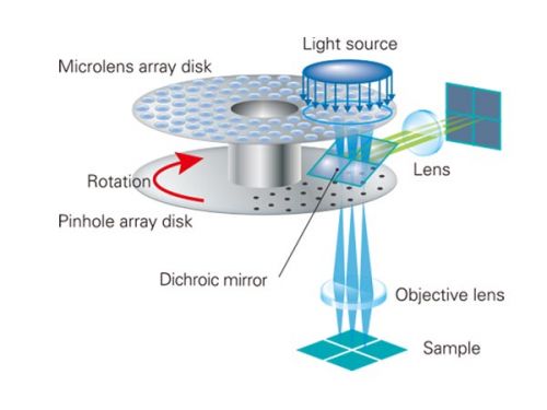
One example of a technological advancement that accelerated the point-by-point collection capacity of laser scanning microscopes is the introduction of a spinning disk. Spinning disk confocal microscopy has to do with the number of pinholes used to block the out-of-focus fluorescent light. While confocal laser scanning microscopy uses a single pinhole, spinning disk confocal microscopy uses an opaque disk with hundreds of pinholes that rotates at high speeds. This allows the entire specimen to be imaged at one time rather than point by point. This can also help to reduce photodamage and photobleaching.
Laser Scanning Microscope Images
As mentioned, the high resolution and contrast of the confocal laser scanning microscope’s images are due to its rejection of unfocused fluorescent light through the pinhole before reaching the detector. After the 3D image is created, observers can manipulate and even explore inside it in certain instances.
Laser Scanning Microscopy vs. Scanning Electron Microscopy
Though similar in name, the main difference between the laser scanning microscope and the scanning electron microscope is the method of illumination for the sample. While laser scanning microscopy uses an excitation laser or lasers, scanning electron microscopy, as its name might suggest, irradiates the sample with a beam of electrons. Reflected electrons or secondary electrons from the surface of the sample produce signals about its topography or composition, which are collected by the detector. As the sample must be in a vacuum, scanning electron microscopy cannot be applied for the observation of live samples.
Laser Scanning Microscope Magnification
Laser scanning microscopes can be used with a variety of magnification objectives. Therefore, the laser scanning microscope’s max magnification depends on the objective being used. Low-magnification objectives are ideal for capturing the structure of an entire tissue sample. If you need to capture the morphology of the cells that make up the tissue, a medium-magnification objective should suffice. High-magnification objectives can then be used to view the microstructure within the cells that make up the tissue.
Sorry, this page is not
available in your country.