Alec De Grand
Meu nome é Alec De Grand e sou gerente de produto de escaneamento de lâminas virtual e microscópios verticais na Olympus. Estou na Olympus há mais de 10 anos, gerenciando produtos clínicos, iniciativas de marketing, cursos de imagem e feiras
Bülent Peker
Olá! Sou Bülent Peker e sou seu especialista designado em microscopia de varredura a laser. Comecei a me interessar por microscopia e fotônica durante meu doutorado em físico-química, onde trabalhei com microscopia de dois fótons resolvida no tempo e a paixão se mantêm até hoje.
Estou na Olympus há mais de 13 anos e ajudei a equipe a lançar nossos microscópios de varredura a laser de última geração no mercado. Estou particularmente fascinado pela aplicação de sistemas multifotônicos e as possibilidades de personalização dos sistemas de escanemaneto a laser.
.jpg?rev=AC43)
Chunsong Yan
Olá, meu nome é Chunsong Yan e sou gerente de desenvolvimento de negócios para ciências da vida na Olympus Austrália e Nova Zelândia. Atualmente sou responsável por sistemas confocal, multifotônico, luz-folha e escaneamento de lâminas. Entrei para a Olympus em 2003, desempenhando várias funções, sempre tentando oferecer a melhor solução Olympus aos nossos clientes.
Daniel Bemmerl
Meu nome é Daniel Bemmerl e estou aqui para ajudá-lo com qualquer coisa relacionada à análise de alto conteúdo 3D e imagens organoides. Entrei para a Olympus como especialista em aplicativos para microscopia TIRF e triagem de alto conteúdo e agora me especializo em software de análise 3D.
Estudei biologia molecular e de desenvolvimento de células-tronco e rapidamente percebi que estava mais interessado nos aspectos mais técnicos da pesquisa e, especialmente, na própria imagem do que nas imagens dos espécimes. Vim para a Olympus Ciências da Vida porque gostei da ideia de trabalhar fora do laboratório e como uma interface entre o cliente e o desenvolvimento. Terei prazer em responder a qualquer uma de suas perguntas sobre imagens 3D e ajudá-lo a melhorar seu fluxo de trabalho de imagem para se concentrar em sua pesquisa.
(2).jpg?rev=9918)
Dr. Nicolas Bourg
Olá! Eu sou o Dr. Nicolas Bourg, diretor técnico (CTO) e cofundador da Abbelight e eu sou o especialista designado para microscopia de localização de molécula única (SMLM) — conhecida como nanoscopia — que inclui técnicas como PALM, dSTORM, SPT-PALM e DNA- PINTURA.
Sou engenheiro optrônico por formação e fiz um doutorado. Doutor em biofotônica na Universidade Paris-Saclay, trabalhando em uma técnica única de nanoscopia 3D com resolução sem precedentes. Com minha equipe de pesquisa, decidimos compartilhar todo o conhecimento que adquiri durante meu doutorado e criamos a Abbelight: uma startup que visa tornar a nanoscopia ainda mais poderosa e totalmente acessível a qualquer biólogo sem treinamento avançado em microscopia. Meu trabalho é responder a todas as suas perguntas sobre nanoscopia, por favor, entre em contato.
Flavio Giacobone
Olá, meu nome é Flavio Giacobone e sou responsável pelas soluções de escaneamento de lâminas nos mercados da Olympus Europa. As imagens sempre estiveram no meu sangue, desde minha graduação em engenharia biomédica, e ingressar na Olympus me permitiu experimentar em primeira mão a mudança para o digital, o que tem acontecido em muitos campos clínicos e de pesquisa. Eu construí minha experiência em imagens de lâminas inteiras a partir do primeiro escâner da Olympus, o dotSlide, então sou uma testemunha ocular de quanto progresso foi feito desde o primeiro produto.
Ganesh Kadasoor
Eu sou Ganesh Kadasoor, associado à Olympus Imagens de alta tecnologia nos últimos 15 anos na Índia. Tenho bacharelado e mestrado em Ciências da Vida e Pós-doutorado Segurado, em Entomologia Médica, pela Universidade de Mysore, Índia. Entrei para a Olympus como especialista Confocal cuidando de todos os sistemas de imagem de alto nível, como sistemas de imagem de células vivas, varredura confocal a laser, TIRF, disco giratório confocal, superresolução e microscópios multi-fótons. Atualmente trabalhando como Gerente Nacional (Suporte de Aplicativos) na Olympus Medical Systems India Ltd, Bangalore, envolvido na condução de programas de formação para Cientistas, Acadêmicos e rede de Distribuidores em todo o país. Também sou responsável por organizar vários workshops de microscopia e imagem, seminários e webinares e dar suporte a cursos de microscopia nacionais e internacionais como tutor/treinador na Índia.
Heiko Gäthje
Olá, meu nome é Heiko Gäthje. Minha experiência em campo amplo, microscopia de fluorescência confocal e processamento de imagem de dados 3D começou quando trabalhava como biólogo — concentrei-me no desenvolvimento neuronal de insetos e na estrutura de proteínas neuronais de ligação de ácido siálico em mamíferos.
Entrei para a Olympus em 2004 e sou instrutor de microscopia na Olympus Academy desde 2008, onde sou responsável pela concepção e introdução de ferramentas de aprendizagem digital. Também dou suporte e conduzo cursos de formação em microscopia no EMBL Heidelberg e no Curso de Inverno de Zurique sobre Microscopia Avançada, onde respondo a muitas perguntas relacionadas ao processamento e análise de imagens.
.jpg?rev=2D93)
Irina Rakotoson
Olá, sou Irina e estou ansiosa para discutir microscopia light sheet com você. Obtive meu mestrado em Biologia Celular, Fisiologia e Patologia com especialização em envelhecimento na Universidade Paris Descartes. Antes de ingressar no PhaseView como Gerente de Produto, trabalhei em um laboratório de pesquisa em Neurociências (SPPIN), otimizando protocolos de limpeza para a instalação central de microscopia.
Junsung Kim
Olá, meu nome é Junsung Kim e sou especialista em microscópio confocal da Olympus Korea. Eu tenho um mestrado em bioengenharia pela Universidade da Coreia. Entrei para a Olympus em 2014 e forneço suporte de aplicativos para clientes com microscópios confocais e multifotônicos da Olympus.
Kathy Lindsley
Eu sou Kathy Lindsley e sou um especialista em aplicativos da Olympus, oferecendo suporte a sistemas de imagem baseados em câmera. Eu tenho um bacharelado em bioquímica pela Universidade do Estado de Iowa. Entrei para a Olympus em 2006 como representante de vendas de imagens de pesquisa e em 2012 fiz a transição para o grupo de aplicações de ciências biológicas. Antes de ingressar na Olympus, trabalhei como assistente de pesquisa em pesquisa acadêmica por 15 anos, ganhando experiência em patch clamp, imagem de cálcio, cultura de tecidos e imunohistoquímica.
.jpg?rev=2E55)
Lauren Alvarenga
Olá! Eu sou Lauren Alvarenga e trabalho como gerente de produto para pesquisa de imagens na Olympus, onde atualmente sou responsável por software de imagem e microscópios invertidos e de superresolução. Eu tenho um bacharelado em comunicações fotográficas biomédicas do Rochester Institute of Technology.
Regulação pós-transcricional do transportador renal de fosfato Npt2a pelo hormônio da paratireóide. Estou na Olympus desde 2015, dando suporte às linhas de produtos FLUOVIEW nos EUA, Canadá e América Latina.

Manoel Veiga
Olá! Meu nome é Manoel Veiga e faço parte da equipe que implementou a aprendizagem profunda no software Olympus. Entrei para a Olympus em 2017, onde desenvolvi expertise em triagem de alto conteúdo, análise de imagem e aprendizado profundo. Também sou um especialista em imagens de fluorescência ao longo da vida.
Comecei a me interessar por análise de dados durante meu doutorado em físico-química — um interesse que desenvolvi depois de descobrir o poder das redes neurais convolucionais e as incríveis tarefas de análise de imagens que elas podem realizar.
Minju Kim
Olá, sou Minju Kim e especialista em microscópio biológico da Olympus Korea. Entrei para a Olympus em 2010 e trabalhei como especialista em vendas e produtos de ponta para células vivas e sistemas de imagem de fluorescência. Além disso, sou encarregado dos programas de treinamento dos distribuidores na Coreia do Sul.
Rebecca Bonfig
Olá! Meu nome é Rebecca Bonfig e sou gerente de produto de microscopia confocal da Olympus. Concluí meu trabalho de graduação na Universidade de Louisville no departamento de fisiologia e biofísica, estudando a regulação pós-transcricional do transportador renal de fosfato Npt2a pelo hormônio da paratireóide. Estou na Olympus desde 2015, dando suporte às linhas de produtos FLUOVIEW nos EUA, Canadá e América Latina.
Shohei Imamura
Meu nome é Shohei Imamura. Sou gerente de projetos estratégicos da Olympus. Tenho quatro anos de experiência em vendas de microscopia científica e sete anos de experiência em planejamento de produto — especialmente software — e gerenciamento e execução de projetos estratégicos. Sou bacharel em comércio pela Universidade Meiji, no Japão.
Srivats Hariharan
Olá! Meu nome é Srivats Hariharan a.k.a Hari e sou gerente de produto da microscopia Confocal, Multifóton e Superresolução na Olympus Singapore. Sou formado em Engenharia Mecânica pela Universidade Tecnológica Nanyang, Cingapura, e tenho experiência em laboratórios de pesquisa biomédica e no A*STAR Microscopy Core Facility, onde dei suporte a pesquisadores em tecnologias de imagem confocal e de células vivas e ajudei a configurar superresolução e microscopia de light sheet de molécula simples.
Entrei para a equipe de ciências biológicas da Olympus Singapore em 2011 como Gerente de Produto e responsável pelo suporte a clientes de pesquisa e parceiros de negócios no Sudeste Asiático e em Taiwan.
.jpg?rev=9E11)
Stefan Marawske
Olá, sou Stefan Marawske e sou seu especialista em microscopia de superresolução. Durante meu doutorado na área de físico-química, criei um microscópio feito em casa para superresolução baseada em localização e rastreamento de partículas. Fiquei fascinado com o fato de que esses métodos são capazes de quebrar o famoso limite de difração de Abbe e foram capazes de resolver estruturas que não podiam ser identificadas antes.
Trabalho para a Olympus há mais de 7 anos e sou o principal responsável por sistemas de imagem de alto nível, como TIRF e disco giratório. Esses sistemas normalmente têm um alto nível de flexibilidade, pois muitos dispositivos diferentes podem ser combinados para várias aplicações, portanto, se precisar de ajuda na definição de sua configuração dedicada, entre em contato.
Takeo Ogama
Sou Takeo Ogama, planejador de produtos sênior e estratégia e gerente de produto para câmeras de microscópio na Olympus. Tenho oito anos de experiência trabalhando no departamento de pesquisa e desenvolvimento em vários produtos — incluindo câmeras — e oito anos de planejamento de produto, marketing e experiência em gerenciamento. Antes de ingressar na Olympus, obtive meu mestrado em física de neutrinos na Universidade de Osaka, no Japão.
Wei Juan Wong
Olá! Meu nome é Wei Juan Wong e sou especialista em produtos para o mercado clínico e de pesquisa. Sou formado em física e tenho experiência de trabalho em um laboratório de pesquisa de biofísica e em uma instalação central de microscopia. Entrei para a equipe Olympus Singapore Ciências da Vida em 2018 como especialista de produto, fornecendo suporte de aplicação para clientes e responsável pelo programa de treinamento para distribuidores na região do Sudeste Asiático.
Klaus Willeke
Hello, I’m Klaus Willeke, Product Marketing Manager at Olympus Life Science Division and I’m responsible for our new X Line objectives. During my geology studies, the polarization microscope was an essential tool for determining and researching mineral compositions and structures. I was always fascinated by how colorful the world of minerals appears through a polarization microscope and how much you can discover with the help of light and good optics.
I’ve been working for Olympus for more than 22 years where I was 17 years a sales representative for industrial and life science microcopy in Germany and after that in European Product Marketing in the Scientific Solutions Division of Olympus, responsible for upright clinical and research microscopes and X Line lenses.
Dr. Hrishikesh Pai
Dr Hrishikesh Pai did his Fellowship in Reproductive Biology from the Royal Women’s Hospital, Melbourne Australia in 1989. He did his Masters in Clinical Embryology and Andrology from the Jones Institute, Eastern Virginia Medical School, USA in 2006. He has won 45 plus awards including the Honorary FRCOG from the Royal College of Obstetricians & Gynecologists, United Kingdom in 2019. Dr Pai has been the Past President of the Indian Society for Assisted Reproduction (ISAR) and is the present Director of Corporate Affairs for International Federation of Fertility Societies (IFFS). Dr Pai is the President Elect of the Federation of Obstetric and Gynecological Societies of India (FOGSI) with a membership strength of 37,000 members. It is the second largest medical body of gynecologists in the world, only second to the ACOG.
.jpg?rev=69FC)
Hiroya Ishihara
Mr. Hiroya Ishihara is an Application Scientist at Olympus. He was studying the epigenetic factors involved in plant regeneration using omics and microscopy in Tokyo University of Science. Confocal and two-photon microscopy were his trusted partner at that time. Therefore, he joined Olympus to make life science more exciting with microscopes. Currently, he is working on a wide range of projects from basic research to product/sales strategy.
Dr. Gowri Balachander
Dr. Gowri Balachander is a Research Fellow at the Yong Loo Lin School of Medicine, National University of Singapore. She completed her PhD at the prestigious Indian Institute of Science, Bangalore, India where she worked on engineering 3D organotypic models for the study of breast cancer metastasis. At NUS, she currently works on morphogenetic models for human liver development and regeneration.
.jpg?rev=AB60)
Joanna Hawryluk
Joanna Hawryluk is a product manager for Olympus Corporation of the Americas located in Waltham, MA. She has been with Olympus since 2017 within the Marketing department specializing in our 3D cell analysis software, lightsheet microscopy, electrophysiology, and our cell culture monitoring system. Dr. Hawryluk received her doctorate degree in Physiology and Neurobiology in 2016 from the University of Connecticut.
.jpeg?rev=F6E8)
Benjamin Compans, Ph.D.
Benjamin received his Ph.D. from the University of Bordeaux. During his Ph.D., in the group of Dr. Choquet at the Interdisciplinary Institute for Neuroscience, he studied the nanoscale organization of glutamate receptors at excitatory synapse and its importance for synaptic plasticity. In 2018, he joined the lab of Professor Burrone at King’s College London for his postdoc where he investigates the molecular organization of inhibitory synapses and its role in regulating neuronal firing, a project for which he obtained a Marie Sklodowska-Curie Action postdoctoral fellowship.
Lin Guo
Lin got his PhD back in 2010 in National University of Singapore working on biophysical research. From 2009 he joined Olympus as Technical and Application specialist taking care of laser based high end imaging system. In 2012, Lin decided to move back to China and taking a position with one of the leading scientific camera manufacturers Photometrics. There, he started as application specialist, later become regional sales manager and finally scientific sales manager for Asia Pacific. In 2021, Lin moved back to Singapore, joining Olympus Singapore as the manager for product and application. Lin has a long experience of various techniques on scientific digital imaging including various camera technologies.
Angela Vasaturo
Hello, my name is Angela Vasaturo, the senior field application scientist at Ultivue. My passion for micro-biology and live-cell imaging began during my post-doc, where I was involved in the early development of multiplex IHC in Europe.
I built my expertise through extensive involvement in post-doc research focused on tumor immunology at the NCMLS in Nijmegen, NL, as well as during my position as Senior Researcher in Dr. Jerome Galon’s Laboratory of Integrative Cancer Immunology at the Cordeliers Research Center.
.png?rev=14CE)
Shogo Usui
My name is Shogo Usui and I'm product leader of CM20 at Olympus Corporation.
For over ten years, I have been an electrical engineer in the life science R&D team in Olympus Tokyo. I hold a Master’s degree in applied physics from the University of Electrical and Communication from Tokyo, Japan.
.jpg?rev=12D2)
Dr. Anne Beghin
Dr. Anne Beghin is a multidisciplinary scientist with fifteen years of extensive research experience across academia and industry. She obtained her PhD in oncology in 2007 at the University Claude Bernard in Lyon (France). She then moved to optical microscopy at the Université de Lyon, where she established the microscopy platform and developed live cell imaging solutions and image analysis services for 4 years.
In 2011, she was recruited by a biotechnology company based in Bordeaux, where she spent 3 years in charge of a tissue analysis service: from biologic samples (whole tissue sections and Tissue Micro Arrays) to image acquisition and analysis with database establishment. She has been part of the Interdisciplinary Institute of NeuroScience IINS for 3 years where she successfully developed a new platform linking the High Content Screening (HCS) approach with super resolution microscopy such as Single Molecule Light Microscopy (HCS-SMLM), a collaboration with pharmaceutical company, Sanofi. Subsequently, she moved to the MechanoBiology Institute (MBI) in Singapore to study organoids using advanced imaging and HCS. This work has resulted in a patent and publications are on-going.
.jpg?rev=339E)
Dr. Xiaotong Cui
Dr. Xiaotong Cui received his B.S. and M.Sc. from the University of Leicester, UK. He went on to receive aPh.D. from the Institute of Life Science, Kyoto University in 2018, where he worked under the direction of Prof. Osamu Takeuchi. From 2018-2020, he was a program-specific researcher in Shenzhen Digital Life Institute and ASHBi, Japan. Dr. Xiaotong has a solid background in molecular cell biology, immunology and he participated in one special program for Key Basic Research of Ministry of Science and Technology, China. Now, he serves as a Field Application Specialist in Bio-Techne China.
.jpg?rev=2687)
Dr. Kasmira Wilson
Dr. Kasmira Wilson is a general surgeon and CSSANZ trainee, having previously completed a BSc(Hons) and MBBS. She is currently undertaking a PhD through the University of Melbourne at Peter MacCallum cancer centre which focuses on translational research in rectal cancer. Her research utilises tumouroid models to explore anti-tumour immunity in rectal cancer in a novel functional cytotoxic assay.
.jpg?rev=74B7)
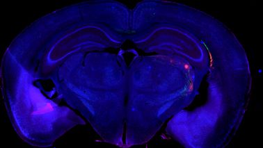
Dr. Dan Zhu
Dr. Dan Zhu is a professor of Huazhong University of Science & Technology, and Vice-Director of Wuhan National Laboratory for Optoelectronics. Her research interests mainly focus on tissue optical clearing imaging and applications. She is the pioneer in the field of in vivo tissue optical clearing, and also developed fast, label-compatible in vitro optical clearing methods. She has authored more than 150 papers including Science Advances, Nature Communications, et al. She is also Fellow of SPIE, and Secretary General & Vice President of Biomedical Photonics Committee of Chinese Optical Society. She serves for journals as editorial member or guest editor, including Biomedical Optics Express, Journal of Biomedical Optics, Scientific Reports, Journal of Innovative Optical Health Sciences, Frontier of Optoelectronics et al.
.jpg?rev=2811)
Dr. Graham Wright
Dr. Graham Wright holds an interdisciplinary PhD in cell biology and physics from The University of Edinburgh and an MBA from the Warwick Business School at The University of Warwick. He is the Acting Director of A*STAR’s Research Support Centre (RSC) and the Director of the A*STAR Microscopy Platform (AMP). RSC offers everything from on-demand access to sophisticated scientific instruments and services, located within varied Technology Platforms, to a research consumables webstore, playing a crucial role in powering biomedical innovation in Singapore.
Dr. Graham has extensive experience, and a strong publication record, in applying advanced light microscopy to a wide range of biomedical research projects. Outside the laboratory, he is committed to science outreach and has featured as a judge on MediaCorp’s National Science Challenge TV show, presented a TEDx talk, had microscopy images displayed on the big screen in Times Square, New York and exhibited work at the National Museum of Singapore.
.jpg?rev=73D5)
Mr. Srivats Hariharan
Mr. Srivats Hariharan is an Applications & Marketing Manager in Olympus life science team in the Asia Pacific region. He holds a bachelor’s degree in Mechanical Engineering from Nanyang Technological University, Singapore and has experience working in biomedical research labs and A*STAR Microscopy Core Facility where he supported researchers on confocal and live cell imaging technologies and help setup single molecule super-resolution and light sheet microscopes. He joined the life-science team of Olympus Singapore in 2011 as Product Manager and in-charge of supporting research customers and business partners in South-East Asia and Taiwan.
.jpg?rev=9660)
Ms. Gency Gunasingh
Ms. Gency Gunasingh completed her Master of biotechnology degree from University of Queensland in 2012 and did her Masters project under Dr. Andrew Prowse and Prof. Peter Gray at Australian Institute for Bioengineering and Nanotechnology. Her primary area of research was large scale generation of cardiac progenitors from embryonic stem cells in 3D. She then worked under Prof. Brian Gabrielli at UQ Diamantina institute on developing 3D tumour spheres in melanoma for in vitro drug testing. She currently works for Prof. Nikolas Haass at UQDI on understanding tumour heterogeneity and tumour architecture in melanoma spheroids.
.jpg?rev=2087)
Dr. Dong Gao
Dr. Dong Gao is the Principal Investigator of Shanghai Institute of Biochemistry and Cell Biology, Chinese Academy of Sciences. His research interests mainly focus on t prostate adult stem cell and the establishment of patients-derived cancer organoid biobank. He has authored more than 40 papers including Cell, Nature Genetics, Cell Stem Cell et al.
.jpg?rev=1436)
Dr. Yu Weimiao
Dr. Yu Weimiao obtained his Ph.D. from the National University of Singapore (NUS) in 2007, majoring in image processing and machine vision. He joined the Agency of Science, Technology and Research (A*STAR) in 2007. He is currently heading Computational Digital Pathology Lab (CMPL) in BII to deepen and extend the R&D with clinical and industrial partners. He is also the joint PI in IMCB leading the Computational & Molecular Pathology Lab (CMPL). His research interests are Computational Biomedical Image Analysis and Quantitative Imaging Informatics. He applied 3D image analysis solution to segment and tracking the cells in 3D to understand the developmental problem and collective cell migration mechanisms. His work was published in Nature Communication, Current biology, Nature Cell Biology, etc. To enhance the applications in clinical diagnosis/prognosis, he co-founded a biotech company, known as A!maginostic Pte. Ltd. He established a world-class joint platform for the immunodiagnosis at the tissue level. The platform allows the researchers, clinicians, and pharma to profile the patient immune signature for diagnosis, prognosis, and drug response study.
.jpg?rev=9A5C)
Dr. Motoki Takagi
Dr. Motoki Takagi received PhD from Graduate School of Agriculture and Life Sciences, the University of Tokyo in 2001. Then he continued his research as a postdoctoral fellow at Institute of Molecular and Cellular Biosciences, the University of Tokyo. He was engaged in the research of nucleic acid drugs at Genencare Research Institute, Co. Ltd. since 2002. Since 2006, he conducted drug discovery research at Biological Systems Control Team, Biomedicinal Information Research Center, Japan Biological Informatics Consortium. Since 2012, he has been researching chemical biology as an associate professor at Fukushima Medical University and became a professor in 2014.
Dr. Ningbo Wu
Dr. Ningbo Wu is an Associate Professor at Shanghai Institute of immunology, Shanghai Jiaotong University School of Medicine. His research interests mainly focus on the function of intestinal stromal cells in intestinal homeostasis and related work was published in Nature and Science CHINA Life Sciences, et al.
Dr. Nuno Costa-Borges
Nuno Costa-Borges is devoted to offering quality control (QC) tests, training, and consulting services to the IVF community worldwide. With over 18 years’ experience, Nuno completed a PhD focused on improving animal cloning efficiency before joining IVI Barcelona as a clinical embryologist. Now, as cofounder and scientific director of Embryotools, Nuno is committed to developing new IVF techniques, which have led to the world’s first babies via maternal spindle transfer for infertility and to the development of the first robotic system for intracytoplasmic sperm injection successfully tested clinically.
Akira Saito
Akira studied veterinary medicine at Tokyo University of Agriculture and Technology, Japan and graduated in 2007. Shortly after, he joined Olympus as application specialist responsible for in vivo imaging systems, high-content analysis systems, and laser confocal systems to support customers in Japan. In 2013, he took over sales promotion for all Olympus life science products. From 2018, he moved to Singapore and joined to support the marketing and application support for the APAC market.
Bob McLean
Bob McLean has over 30 years’ experience as a microbiologist, during which time he and his lab have done a number of studies on surface-adherent microorganisms (biofilms). In 1998, he and his colleagues had an experiment on the space shuttle with John Glenn, in which they were one of the first research groups to show that biofilms could form in microgravity. Since that discovery, there have been a number of biofilm issues, notably instances of fouling in the water recovery system in the International Space Station and other spacecraft. In 2015, Bob, along with collaborators at Arizona State and the Johnson Space Center, received a NASA grant to study biofilm formation during spaceflight. Confocal and other types of microscopy have been instrumental in these investigations.
Jesse Chao
Jesse completed his Ph.D. at the University of British Columbia (UBC) in cell biology and genomics. He then continued his training at the University of California, San Diego. After, he switched his focus to developing machine learning approaches for assessing the physiological impacts of genetic variants associated with hereditary cancer at UBC. During this time, he started to develop deep learning approaches to automated phenotypic profiling based on high-content imaging data.
James Lopez
James Lopez received his Ph.D. in biomedical sciences from the University of Chicago in 2010. With nearly a decade of experience in calcium imaging, FRET, live cell imaging, and intravital imaging, James joined Olympus as a confocal and multiphoton sales representative. He later transitioned to the Olympus Life Science Applications Group, supporting confocal and multiphoton systems. Now he manages the Life Science Applications Group in the US, Canada, and Latin America markets.

Yosuke Yoneyama
Yosuke Yoneyama obtained his Ph.D. from The University of Tokyo in Japan, where he continued his post-doctoral work on insulin/insulin-like growth factor signaling with a focus on spatiotemporal control of the intracellular signaling system in the laboratory of Dr. Shin-Ichiro Takahashi. He then joined the laboratory of Dr. Takanori Takebe at Tokyo Medical and Dental University in Japan as an assistant professor. He now focuses on human organoids, in particular liver organoids, that are derived from stem cells, including induced pluripotent stem cells (iPSCs), to reconstitute multiple lineages of liver cells both for human development and modeling diseases such as non-alcoholic steatohepatitis.
Ewa Goldys
Professor Ewa M. Goldys is Deputy Director of the Australian Research Council Centre of Excellence in Nanoscale Biophotonics (cnbp.org.au) and Professor at the Graduate School of Biomedical Engineering, the University of New South Wales, Sydney, Australia. She is Fellow of SPIE, OSA, the Australian Academy of Technological Science and Engineering (ATSE), and winner of the 2016 Australian Museum Eureka Prize for ‘Innovative Use of Technology.’ She has ongoing involvement with SPIE BIOS, the world's largest international biomedical optics meeting and SPIE's Photonics West where she serves as Track Chair in Nanobiophotonics.
Her research spans the areas of biomedical science, bioimaging, biosensing, and materials science. She developed novel approaches to biochemical and medical sensing and deployable medical diagnostics. Current projects focus on cancer nanotechnology and non-invasive high-content imaging of colors and patterns in cells and tissues.
Laura Vittadello
Dr. Laura Vittadello is working as a post-doc in the physics department of the Osnabrück University in the ultrafast physics research group. Her research focus is on the fundamental study and application of a new type of marker, harmonic nanoparticles, that are specially designed for biological applications that involve nonlinear microscopy.

Francesco Cardarelli
After receiving his M.Sc. in Biological Sciences from the University of Pisa in Oct 2003 and his Diploma in Biological Sciences in the same year (both with honors) from SNS, Francesco Cardarelli worked at the NEST Laboratory of SNS as a Ph.D. student in Molecular Biophysics under the supervision of Prof. Fabio Beltram. He started his interdisciplinary research at the crossroads between cell biology and physics, using advanced fluorescence microscopy methods to study the intracellular transport properties of virus-derived peptide sequences. After graduating, he became a Post-Doctoral Fellow at the Laboratory for Fluorescence Dynamics, University of California at Irvine, under the supervision of Prof. Enrico Gratton, where he coordinated the research activity for the development of new spatial variants of fluorescence correlation spectroscopy to detect barriers to molecular diffusion/flow in live cells. In Dec 2010 he was hired by the CNI@NEST (IIT) as a Post-Doctoral Fellow. Back in Italy, he started working to develop new fluorescence-based imaging and analysis methods to study single molecules in complex biological systems with high spatiotemporal resolution. This research was boosted by a number of funded grants (and established collaborations) and by an independent scientific position, first at CNR as a Researcher, then at SNS as Professor in Applied Physics.
The focus of his research is on the development of new optical microscopy techniques to increase the amount of quantitative information that can be extracted from investigations on living matter. For instance, in recent years, he and his team introduced a number of new spatiotemporal fluctuation-analysis tools (iMSD, iRICS, nD-pCF, diffusion tensor analysis, etc.) to extract structural and dynamic properties of biological objects, from molecules to entire sub-cellular structures, in their complex natural environment. Such a toolbox is becoming a new paradigm for biophysical investigations at the nanoscale, as featured in the “New and Notable” section of Biophysical Journal (2016 Aug 23; 111(4): 677–678). In 2014, together with his Team, they demonstrated the occurrence of short-range protein Brownian motion in the cell cytoplasm, being among the first to challenge the current view of the structural organization of the crowded intracellular environment. Finally, by combining this toolbox with feedback-based orbital tracking, they demonstrated that even the nanoscopic and dynamic environment of intracellular organelles can be quantitatively probed.
Sandrine Roy
Dr. Roy completed a double-major in Biochemistry and Microbiology followed by the completion of a Doctor of Philosophy in 2002 in the field of Molecular Biology/Cell Biology at the University of Queensland Australia. She travelled abroad to undertake a post-doctoral position at Washington University in St Louis, USA. She then returned to Australia to continue her post-doctoral studies.
With her extensive microscopy experience, she was appointed as the University of Queensland Diamantina Institute Microscopy Facility Manager in 2009 and later as manager of microscopy services at the Translational Research Institute in Brisbane until 2019.
She is now Business Development Manager at Olympus Australia, where her experience and knowledge is used to support customers both in Australia and New Zealand.

Seungil Kim
Seungil Kim, Ph.D., is a Staff Scientist and Microscopy Team Manager at the Lawrence J. Ellison Institute for Transformative Medicine at USC. Dr. Kim completed his B.S. and M.S. degrees in South Korea. He then moved to Washington University and received a doctoral degree in Developmental Biology. He carried out his postdoctoral research in the department of Cell and Tissue Biology at UCSF. Seungil has over 10 years of experience working with various in vitro/in vivo models and advanced cellular imaging techniques. His current research focus is to understand the contributions of the tumor microenvironment to drug response, using patient-derived 3D organoids as a model system. Moreover, he is developing high-throughput automated imaging methods to screen novel drug compounds in colorectal cancer.
Alfonso J. Schmidt
Alfonso has a decade of experience working in a shared resource lab (SRL) with a vast knowledge in histology, fluorescent microscopy, and image analysis. His work has been focused in maximizing the capabilities of the equipment available and in creating technical protocols and training modules for the scientific community. Currently, Alfonso oversees the Histology and Bioimaging Facility as part of the Hugh Green Cytometry Centre (HGCC) at the Malaghan Institute of Medical Research in Wellington, New Zealand.

Tong Wu
Tong Wu joined Olympus in 2012 after completing her Ph.D. in China (State Key Laboratory of Fine Chemicals, DLUT). Now, Tong is a business development manager, supporting high-end microscopes in Olympus Australia. With a research background in fluorescent dyes for bio-imaging and bio-labelling, Tong Wu is enthusiastic to engage with customers’ research applications.
Ruben Portugues
Prof. Portugues is a neurobiologist studying sensorimotor control. His research group uses behavior, modeling, optogenetics, in vivo electrophysiology, and whole brain functional calcium imaging to dissect learning, memory, and action selection in the larval zebrafish.
Prof. Portugues studied mathematics and did his Ph.D. in theoretical physics at Trinity College in the University of Cambridge. After a short postdoctoral fellowship in physics at Centro de Estudios Cientificos in Valdivia, Chile, he joined Professor Florian Engert’s laboratory at Harvard University and switched research interests to neuroscience. In 2014 he was appointed Max Planck Research Group Leader at the Max Planck Institute of Neurobiology in Martinsried. Since 2020 Prof. Portugues is an assistant professor at the TUM.
.jpg?rev=EF4C)
Dr. Thomas Bauer-Jazayeri
Atsuya Toda
Atsuya Toda is an assistant manager for Global Life Science Marketing at Evident. He has more than fifteen years’ experience in life science microscopy sales, sales planning, and marketing in Japan. In 2021, he moved to the Global Life Science Marketing team where he is the marketing representative for the APEXVIEW APX100 all-in-one microscope. He holds a Bachelor of Economics degree from Doshisha University, Japan.
Dr. Laura Lleras Forero
Laura Lleras Forero is the product marketing manager for cell culture products at Evident. She completed her PhD at King’s College London and undertook postdocs in Utrecht, Berlin, and Münster. She has been with Evident since 2021, supporting cell culture microscopy solutions, the SLIDEVIEW™ VS200 research slide scanner, and the APEXVIEW™ APX100 all-in-one microscope for Europe, the Middle East, and Africa (EMEA).
Ines Hartmann
Ines Hartmann is an application specialist for cell handling at the Eppendorf headquarters in Hamburg, Germany. She joined the company in 2008 and has worked on several topics in cell handling, liquid handling, and consumables. She obtained her diploma degree in biology at the Humboldt University of Berlin, Germany and has expertise in various laboratory techniques.
Amin El-Heliebi
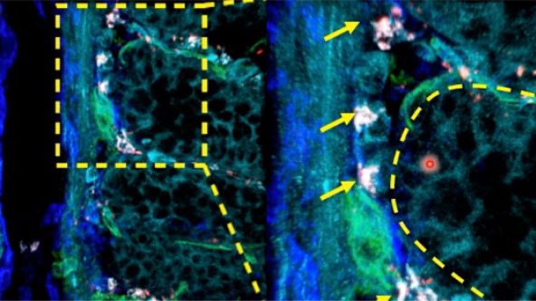
Featured Speaker Amin El-Heliebi (Professor) Professor Amin El-Heliebi was educated in Graz, Stockholm and Buenos Aires and was appointed Research Professor 2021 at the Medical University of Graz, Austria. His research focus lies in molecular biomarkers, cancer research, liquid biopsy, tumor biology, and spatial transcriptomics. His overarching research question deals with understanding tumor dissemination. His research group investigates liquid biopsies, such as circulating tumor cells (CTCs) and circulating tumor DNA (ctDNA). Their overall aim is to track and trace liquid biopsies back to the originating tumor mass and identify the mechanisms involved in spreading of metastatic disease.
Pergunte a um especialista:
Investigating Tumor Dissemination by Spatial Transcriptomics

Transforming Precision Imaging: Meet the FLUOVIEW™ FV4000 Confocal Microscope

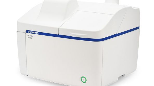
Introduction to the APEXVIEW™ APX100 Digital Imaging System

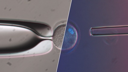
How Polarized Light Can Assist Embryologists in Clinical Routines

Unveiling Nanoscopic Realms: A Journey into Super-Resolution Microscopy
Multiplexing and Deep Tissue Imaging with NIR Confocal Laser Scanning Microscopy (Encore Edition)
Good Cell Culture Practice—How to Improve the Reproducibility of Your Experiments

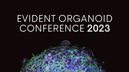
EVIDENT Organoid Conference 2023 - Looking Deeper, Capturing Complexities
Exceptional Imaging Made Easy: Meet the APEXVIEW™ APX100 All-in-One Microscope


Technology Evaluation: Deciphering Cell-Cell Interactions in a 3D Microenvironment at a Single-Cell Resolution

Conferência de Bioimagem da Olympus: Explorando Novas Dimensões | Evento virtual de 3 dias | De 9 a 11 de março de 2022
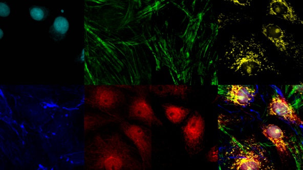
Imagem de super-resolução de célula viva: O grande retrato das coisas pequenas


FV3000 Red Near-Infrared (NIR) Solutions for Confocal Microscopy | 2 p.m.


FV3000 Red Near-Infrared (NIR) Solutions for Confocal Microscopy | 10 a.m.
Olympus Organoid Conference 2021
.jpg?rev=3E0D)
Objetivas de microscópio: onde a mágica acontece

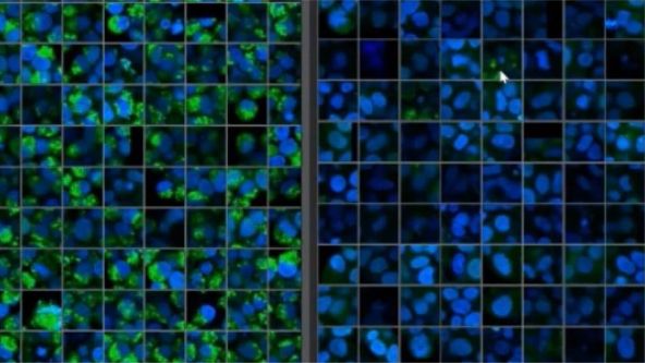
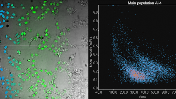
Triagem de alto conteúdo: análise personalizada simplificada


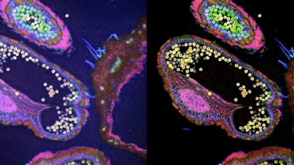
Análise tridimensional de imagem de alta produtividade operacional de organoides e esferoides derivados do paciente.

Olympus Discovery Summit: Advance Your Imaging | 26 e 27 de outubro de 2021
.jpg?rev=1940)
Processamento digital de imagens – parte 2: filtro de processamento de imagens avançado

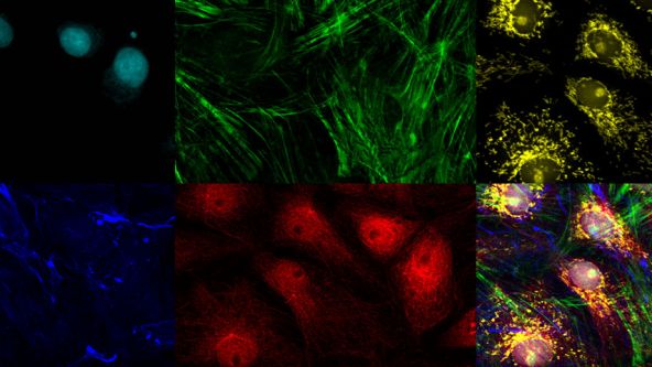
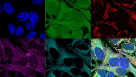
Obtenção de imagens de múltiplas estruturas subcelulares em escala nanométrica em 3D

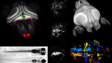
Whole-Brain Functional Calcium Imaging Using Light Sheet Microscopy

Product Demo: SLIDEVIEW™ VS200 Research Slide Scanner

Product Demo: SLIDEVIEW™ VS200 Research Slide Scanner

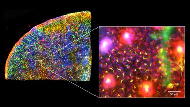
The Use of Multiplexing in Microscopy for Better Understanding the Skin Immune System in the Context of the Tissue


Recent Advances in 3D Imaging and AI-Driven Data Analysis

Now You Have the Power to See More

Metabolic Imaging in Langerhans Human Islets with MPE and FLIM

Product Demo: IXplore™ SpinSR Confocal Super Resolution System
In-Vivo Tracking of Harmonic Nanoparticles by Means of a TIGER Widefield Microscope

Hyperspectral and Brightfield Imaging Combined with Deep Learning Uncover Hidden Regularities of Colors and Patterns in Cells and Tissues

Product Demo: FLUOVIEW™ FV3000 Confocal Laser Scanning Microscope
Product Demo: FLUOVIEW™ FV3000 Confocal Laser Scanning Microscope
Evolution of Scientific Digital Imaging Technologies and their Applications

Deep Learning Approaches to Automated Phenotypic Profiling

Deconvolution of 3D Image Stacks
Confocal Microscopy and Its Use for a Spaceflight Experiment

Accelerating Image Analysis with TruAI™ Deep Learning Technology
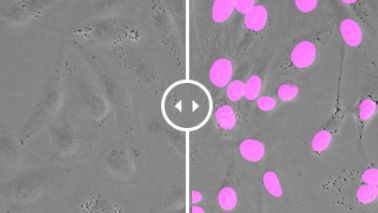
A New Way of Thinking—Object Detection with Deep Learning
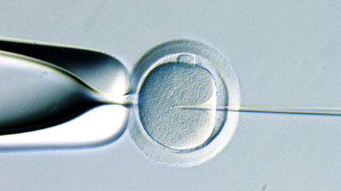
ICSI - How to improve your technique
NoviSight™ Demonstration: 3D Image Analysis and Statistical Software for Organoids and Spheroids
Study the Function of Stromal Cells through Intestinal Organoid Co-Culture Technology

An In Vitro System for Evaluating Anticancer Drugs Using Patient-Derived Tumor Organoids

3D Segmentation for Fluorescence Images: From Qualitative to Quantitative

Prostate Cell Lineage Hierarchy and Plasticity

Investigating Spheroid Architecture Using the FV3000 Confocal Microscope

Advances in 3D Optical Imaging Technologies: An Overview

3D Microscopy: Understanding the Give and Take on Instrument Performance to Enable Informed Decisions

Tissue Optical Clearing Imaging: From In Vitro to In Vivo

Utilizing Tumoroids to Explore Anti-Tumor Immunity in Rectal Cancer

Converting from 2D to 3D: Bio-Techne Solutions for Your 3D Culture

Culture and Quantitative 3D Imaging of Organoids: Challenges and Solutions

A Smarter Approach to Culturing and Nurturing Your Cells

Modern Slide Scanning: Single-cell Phenotyping on Fixed Samples (Encore Edition)


.jpg?rev=6A21)
To the Diffraction Limit and Beyond: The Nanoscale Organization of Axo-Axonic Synapses | 2 p.m.

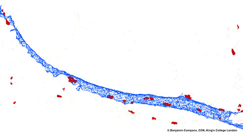
To the Diffraction Limit and Beyond: The Nanoscale Organization of Axo-Axonic Synapses | 10 a.m.
Light Sheet Microscopy – New multi-resolution and -color imaging methods

Olympus Organoid Conference: Think Deep, See Deeper | 3-Day Virtual Event | September 7-9, 2021
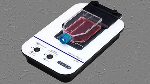
Create a Smarter Cell Culture Workflow


Digital Image Processing: Point and Local Operation Filters (Encore Edition)
Depth Matters: Transforming Biology for More Realistic and Meaningful Pursuits

.jpg?rev=FD74)
Microscope Objectives—Where the Magic Happens (Encore Edition)
Microscopia light sheet para uma análise mais profunda sobre a vida
FluidFM: uma nova abordagem para edição de genes CRISPR por entrega intranuclear direta
Ensaios 3D: software inteligente, análises perspicazes

Multiplexação e imagens de tecidos profundos com microscopia de varredura a laser confocal no infravermelho próximo


Escaneamento de lâminas moderno: fenotipagem de célula única em amostras fixas
Aprendizado profundo: abrindo portas para novos aplicativos

Olympus Discovery Summit—Looking Forward: A New Era of Research
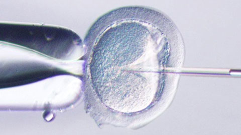
ICSI—Past, Present & Future

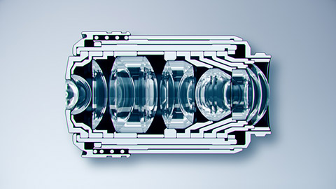
Microscope Objectives—Where the Magic Happens

Think Deep, See Deeper with Near-Infrared Laser Scanning Confocal Microscope

Digital Image Processing: Point and Local Operation Filters
