
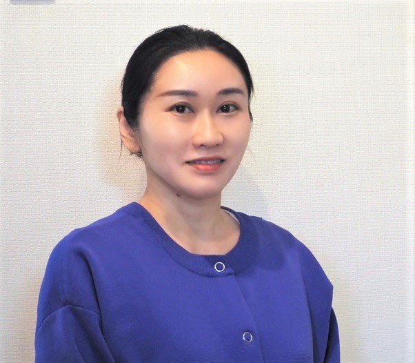
Takeo Ogama
Takeo Ogama是Evident高级产品与策略规划师,也是显微镜相机产品经理。他拥有8年的各种产品研发部门(包括相机部门)的工作经验,以及8年的产品规划、营销和管理经验。他拥有日本大阪大学中微子物理学硕士学位。
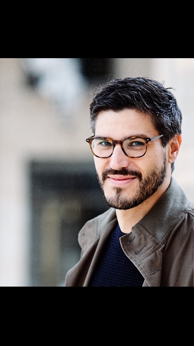
Alket Mertiri
Alket Mertiri博士拥有光学和光子学背景。他在波士顿大学获得了材料科学与工程学博士学位,期间,他致力于开发新型显微镜技术。2018-2019年,他在奥林巴斯担任助理产品经理,通过寻找高级技术(从基础显微镜到全内反射显微镜)解决方案推进科学家的研究。
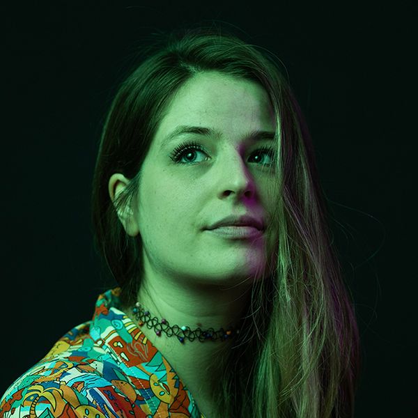
Lauren Alvarenga
Lauren Alvarenga是Evident临床显微镜高级产品经理。她的专业涉及物镜和成像软件。她拥有罗切斯特理工学院生物医学摄影传播专业理学士学位。

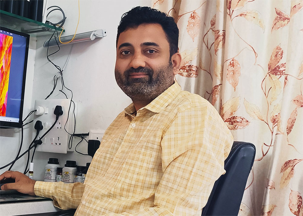
Ralf Schaefer
Ralf Schaefer毕业于汉堡大学,获得了化学专业学位。他曾在多家机构和公司的各种技术环境中从事营销传播工作。Ralf在奥林巴斯的不同项目中以及不同岗位上都塑造并提升了公司显微镜和行业业务的知名度。
Kerry Israel
Kerry Israel是奥林巴斯美洲公司科学解决方案部门生命科学营销与沟通经理。她拥有布兰代斯大学文学学士学位,并且在从广告和社交媒体策略到基层推广的市场营销各个层面拥有超过15年的工作经验。

Alec De Grand
Alec De Grand是Evident虚拟玻片扫描和生命科学正置显微镜产品经理。他在Evident工作超过10年,在此期间,他负责过临床产品、营销活动、成像课程和行业展会的管理工作。
Carlo Alonzo
Carlo Alonzo博士是Evident生命科学显微镜部销售代理。他帮助科学家确定并理解支持其研究目标的技术。由于关注生物医学光学,他加入了波士顿学术界,并在那里花了几年时间深入研究多光子显微镜的使用。他拥有菲律宾大学物理学博士学位,并在丹麦技术大学和哈佛大学医学院麻省总医院接受了博士后培训。

Joanna Hawryluk
Joanna Hawryluk博士是Evident研究用成像产品经理。她目前负责我们的细胞培养孵化监控系统、3D细胞分析软件、电生理学显微镜和光片显微镜。Joanna于康涅狄格大学生理学和神经生物学系获得博士学位。

Laz Amador
Laz Amador是Evident国家应用团队高级经理。Laz拥有36年显微镜销售经验,自1988年以来一直在Evident工作。在加入应用团队之前,Laz曾担任拉丁美洲公司的技术营销执行董事。Laz与他的妻子Jan住在佛罗里达州的韦斯顿。
Rebecca Chandler
Rebecca是奥林巴斯科学解决方案公司(Olympus Scientific Solutions)的特约撰稿人。她拥有恩迪科特学院的新闻学学士学位,擅长撰写与科学和工业领域趋势和技术相关的文章。Rebecca与奥林巴斯工程师和科学家密切合作,负责撰写有关最新激光扫描、超分辨率、多光子、正置、体视和倒置显微镜系统以及前沿光学部件、相机和软件的文章。跟进她的作品即可了解奥林巴斯在细胞学、病理学、教育等众多领域的最新应用。

JamesLopez博士
James Lopez于2010年在芝加哥大学获得了生物医学科学博士学位。James在钙成像、荧光共振能量转移、活细胞成像和活体成像方面拥有近十年的经验,他加入Evident时担任共焦和多光子销售代理。后来,他被调到Evident生命科学应用团队,从事共焦和多光子系统的支持工作。现在,他负责管理生命科学应用组在美国、加拿大和拉丁美洲市场的运营。
.jpg?rev=29C8)
Matthew Weitzman
Matthew Weitzman拥有特拉华大学生物科学博士学位,并拥有多年的高级成像经验。2014-2021年,他在奥林巴斯从事共焦和多光子成像系统工作,为产品开发和应用支持提供协助。
Janeen Manning
Janeen Manning是Evident临床和教育显微镜产品经理。在加入Evident前,她曾在一家专门生产传染病医疗设备的公司工作。她拥有缅因大学的生物化学、微生物学和分子生物学理学学士学位,以及哈佛大学生物技术文科硕士学位。
Christopher D. Higgins
Christopher Higgins是Evident生命科学部业务发展经理。他在显微镜领域拥有24年丰富的工作经验,主要从事各种生物光学显微镜产品管理和应用(研究和临床应用)方面的工作。
Chris充分利用目前职位,直接与研究人员和病理学家合作开发新技术和新应用。利用当前虚拟显微镜系统拍摄的图像不仅能够对单个细胞的情况进行描述,还能够对整个组织的情况进行描述,在研究、教育以及其他方面都发挥着重要的作用。Chris认为临床领域正逐步接受更广泛的全玻片成像和人工智能分析应用,因此,下一个重大前沿工作就是为全玻片成像和人工智能分析向新时代迈进做好准备。
Chris拥有迈阿密大学学士学位,是数字病理学协会成员。他是一名狂热的户外运动爱好者,也是一位优秀的自然摄影师,目前居住在美国宾夕法尼亚州的Lehigh Valley。

Cheng-Hao Chien
Cheng-HaoChien博士在台湾阳明大学获得生物光子学博士学位,并在波士顿塔夫茨大学医学院神经科学系接受了博士后培训。他在高级显微镜和生命科学研究方面有十年经验。2020-2021年,他在奥林巴斯从事多光子显微镜和定制解决方案方面的工作,提供产品和应用支持。
Brendan Brinkman
Brendan Brinkman在干细胞领域(包括小鼠原代神经球的分离)工作多年。几年后,他加入奥林巴斯,担任核心成像设施经理,并协助建立新型系统。2017年,他结束了在奥林巴斯东京总部为期两年半的工作。在东京期间,他帮助开发了下一代解决方案。
Kathy Lindsley

Manoel Veiga
Manoel Veiga于圣地亚哥·德·孔波斯特拉大学获得物理化学博士学位,从事皮秒和飞秒时间分辨光谱的研究。在马德里康普顿斯大学和明斯特大学完成两个博士后研究后,他加入PicoQuant担任高级科学家,从事时间分辨光谱、荧光寿命成像显微镜(FLIM)和荧光相关光谱(FCS)领域的研究。现在,Manoel在Evident德国公司担任全球应用专员,侧重于高内涵分析(HCA)和深度学习。
Heiko Gäthje
Rebecca Bonfig
Rebecca Bonfig博士在路易斯维尔大学生理学和生物物理学系完成了研究生学习,研究内容为甲状旁腺激素对肾脏磷酸盐转运体Npt2a的转录后调节。2015年至2022年,Bonfig博士在奥林巴斯从事共焦显微镜工作,作为产品经理为FLUOVIEW系列产品提供支持。
Bülent Peker
Flavio Giacobone
Shohei Imamura
Shohei Imamura是Evident策略项目经理。他拥有四年的科研显微观察销售工作经验以及七年的产品规划、策略项目管理和项目执行经验。他拥有日本明治大学商学学士学位。
Daniel Bemmerl
Paul Monnier
GulpreetKaur博士
GulpreetKaur博士在威斯康星大学麦迪逊分校获得了分子生物学博士学位,在学习期间,她利用活细胞转盘共聚焦显微镜进行细胞器生物学研究。
Kaur博士在显微镜方面拥有超过六年的经验,并从事过各种技术工作,包括共焦、转盘式、超分辨率和光片成像。她曾担任威斯康星大学麦迪逊分校显微镜和图像分析核心设施的培训师,并为科学家提供实验设计和成像条件优化方面的咨询。
2018年至2022年,Kaur博士帮助科学家在他们的研究项目中应用奥林巴斯显微镜和成像技术。

Nicolas Bourg博士
Sarah Williams
在2013年加入奥林巴斯营销沟通部门之前,Sarah曾在广播媒体行业担任近十年的研究员和撰稿人。目前,Sarah将自己身为作家和编辑的技能运用到为奥林巴斯产品及专业领域相关主题提供引人入胜的高质量素材上。她曾撰写过包括超声(UT)和涡流检测(ECT)、相控阵(PA)、全聚焦方法(TFM)和全矩阵采集(FMC)、以及工业显微镜和内窥镜等一些无损检测(NDT)技术。她经常调研奥林巴斯产品对改善我们身边的世界的品质和安全所做出的贡献。Sarah在魁北克市的办公室工作,并与伴侣大卫(David)和三个孩子Sophie、Anouk和Éloi住在一起。
MikeWoerdemann博士
Mike Woerdemann在基于结构光场的光学镊子专业技术领域获得了博士学位。多年以来,他曾担任应用专员为高内涵筛选系统提供支持,在此期间,他在该领域获得了深刻、实用的知识。现在,Mike担任EVIDENT欧洲专业技术中心高内涵筛选站和成熟深度学习产品的产品经理。

Chunsong Yan
Chunsong Yan是Evident澳大利亚和新西兰公司生命科学部业务发展经理。他目前负责共焦、多光子、灯片和玻片扫描系统。自加入Evident以来,Chunsong虽然担任过不同的职位,但他一直努力为客户提供更好的成像解决方案。
Minju Kim
Junsung Kim
Ganesh Kadasoor
Wei Juan Wong
Wei Juan Wong是Evident数字玻片扫描系统应用专员。她开始在新加坡担任产品专员,为使用宽场显微镜(包括SLIDEVIEW VS200研究用玻片扫查器)的东南亚客户提供支持。后来,她来到德国,加入EVIDENT欧洲专业技术中心,担任数字玻片扫描系统应用专员,为全球客户提供应用和营销支持。Wei Juan拥有物理学学位,曾在生物物理研究实验室以及显微镜核心设施工作。
Srivats Hariharan
Stefan Marawske
Bunryu Arashi
Bunryu Arashi在Evident欧洲公司担任原始设备制造商经理。Bunryu在显微镜产品开发方面拥有12年经验,他曾为BX和CKX显微镜系列产品设计物镜和相机转换器,以及照明光学系统。他拥有日本大阪大学工程学硕士学位。

Craig Rappaport
Craig Rappaport是Evident生命科学部显微镜高级销售代理。他拥有26年的显微镜销售经验,以及十几年的科技营销经验。自加入Evident以来,Craig一直从事市场分析、客户需求调查和新产品研究。他自2012年起加入销售团队,服务范围涵盖南加州的圣地亚哥和Imperial Counties。Craig在加州大学圣巴巴拉分校获得文科学士学位,在杜克大学福库商学院(Duke University Fuqua School of Business)获得MBA学位。
Marcus Krauel
Marcus Krauel在Evident欧洲公司担任原始设备制造商经理。他运用自己在光学领域12年的工作经验,在工业到医疗等各种应用方面为我们欧洲的原始设备制造商客户提供帮助。Marcus拥有物理学和光学工程学位。
Klaus Willeke
Klaus Willeke是Evident生命科学部的一名产品营销经理。Klaus在Evident的工作时间超过22年。刚开始的17年间,他在德国担任工业和生命科学显微镜销售代理,后来,他加入欧洲产品营销团队,负责正置临床和科研用显微镜以及X系列物镜相关工作。
Yu Kikuchi
Yu Kikuchi是Evident元器件产品和显微镜产品光学工程师。他拥有显微镜产品光学设计和评价经验。Yu专注于开发定制产品,负责匹配技术规格与客户要求,以及现场测试和校直。他拥有日本东北大学分子生物学硕士学位。
Stephan Fliegel
Stephan Fliegel于2007年在明斯特应用科学大学获得物理工程专业学位,专注于激光应用技术。他刚开始担任应用专员,然后在工业激光器领域担任产品和项目经理,专注于客户专用激光系统的布局设计、开发和规划。现在,他担任Evident光学显微镜相机产品经理。

DanielGöttel博士
Daniel Göttel在德国海德堡的德国癌症研究中心获得人类遗传学博士学位。多年以来,他曾担任虚拟玻片扫描系统应用专员,并在该领域获得了深刻、实用的知识。现在,Daniel担任Evident SLIDEVIEW VS200研究用玻片扫查器和Net Image Server(NIS)SQL销售支持经理。
Lee Botes
Lee Botes拥有24年的奥林巴斯显微镜销售经验。Lee具有微生物学背景,她的显微镜销售经历始于南非。Lee是Evident新西兰销售团队成员,负责惠灵顿及南岛的工业、临床和科研用显微镜业务,以及新西兰地区的共焦显微镜和玻片扫查器业务。
Masato Yabe
Masato Yabe就职于Evident光学开发部,参与显微镜产品开发已有19年,他为物镜和自动聚焦装置设计光学系统。他拥有日本和歌山大学工程学硕士学位。
Shodai Hosono
Shodai Hosono是Evident机械工程部门成员。他参与开发显微镜产品已逾十年。作为一名机械工程师,他曾设计过各种显微镜和模块化显微镜组件。他拥有日本埼玉大学工程学学士学位。
Kei Sato
Kei Sato在Evident电气工程部门工作,从事显微镜产品开发已有9年。作为一名产品负责人,Kei负责工业显微镜和显微镜元器件的电气设计。
Joanna Hawryluk
Joanna Hawryluk博士是Evident研究用成像产品经理。她目前负责我们的细胞培养孵化监控系统、3D细胞分析软件、电生理学显微镜和光片显微镜。Joanna于康涅狄格大学生理学和神经生物学系获得博士学位。

Dr. Hunter Hines
Dr. Hunter Hines received his PhD in 2019 while registered in a joint international collaboration with Bournemouth University (UK) and Harbor Branch Oceanographic Institute (HBOI) at Florida Atlantic University. He conducted research into microbial communities found in tropical aquatic and terrestrial ecosystems in Florida, focusing on the biogeography and biodiversity of ciliates.
Today, Dr. Hines continues his research at HBOI, where he examines microbial communities and the impact they have on ecosystems. He is also the senior scientist on the Amoray Marine Conservation Team at the Amoray SCUBA Resort in Key Largo, Florida.
On social media, Dr. Hines plays a significant science outreach role for microbiology and ecology. During his PhD, he created the Instagram account Microbialecology, which has over 140,000 followers. He is a frequent collaborator with the popular YouTube channel Brave Wilderness, assisting with everything microbial and featuring in several episodes.
Kenji Ono
Kenji Ono在Evident光学元器件部门工作。自加入公司以来,他在我们的成像业务中帮助进行数码相机光学系统的规划和开发。现在,他负责原始设备制造商显微镜营销活动,将自己的知识应用于光学领域。Kenji于日本北里大学获得学士学位。
Kristina Mayer
Kristina Mayer是Evident欧洲产品营销团队的倒置显微镜产品营销经理。她在Evident工作超过10年,负责IXplore倒置显微镜系列产品相关工作,为研究用成像(包括欧洲、中东和非洲地区的全内反射荧光显微镜(TIRF)和转盘共聚焦显微镜)提供支持。Kristina拥有不来梅雅各布斯大学细胞生物学博士学位。
Benjamin Compans, Ph.D.
Yuan Yue
Yuan Yue在Evident市场策略部工作,主要负责数字营销。她在Evident有近6年的营销传播经验。
Shogo Usui
Dr. Anne Beghin

Dr. Xiaotong Cui

Dr. Kasmira Wilson

Dr. Dan Zhu

Dr. Graham Wright

Mr. Srivats Hariharan

Ms. Gency Gunasingh
Dr. Dong Gao

Dr. Yu Weimiao
Dr. Motoki Takagi
Dr. Ningbo Wu
.jpg?rev=C8C3)
Mr. Hiroya Ishihara

Marie Kawasaki
Marie Kawasaki就职于Evident生命科学营销部门工作。她从事临床研究相关显微镜的营销已有7年,目前负责生育治疗研究(卵胞浆内单精子注射)行业的全球销售推广。
Wei Juan Wong
Wei Juan Wong是Evident数字玻片扫描系统应用专员。她开始在新加坡担任产品专员,为使用宽场显微镜(包括SLIDEVIEW VS200研究用玻片扫查器)的东南亚客户提供支持。后来,她来到德国,加入EVIDENT欧洲专业技术中心,担任数字玻片扫描系统应用专员,为全球客户提供应用和营销支持。Wei Juan拥有物理学学位,曾在生物物理研究实验室以及显微镜核心设施工作。
Samuel M Lawrence
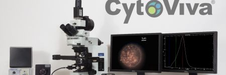
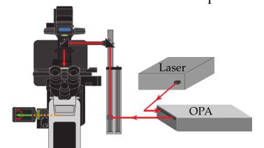
Samuel M. Lawrence is the chief executive officer and cofounder of CytoViva, Inc. He led the development of the core, patented optical illumination technology that is the foundation of CytoViva’s products, as well as played a key role in the development of the patented CytoViva 3D imaging technology. He continues to lead all product development efforts and actively supports the company’s sales efforts both domestically and internationally.
Manoel Veiga
Manoel Veiga于圣地亚哥·德·孔波斯特拉大学获得物理化学博士学位,从事皮秒和飞秒时间分辨光谱的研究。在马德里康普顿斯大学和明斯特大学完成两个博士后研究后,他加入PicoQuant担任高级科学家,从事时间分辨光谱、荧光寿命成像显微镜(FLIM)和荧光相关光谱(FCS)领域的研究。现在,Manoel在Evident德国公司担任全球应用专员,侧重于高内涵分析(HCA)和深度学习。
ManfredKässens博士
ManfredKässens博士主导EVIDENT欧洲专业技术中心技术产品信息部,同时也是Evident全球营销传播部成员。他拥有德国明斯特大学物理学博士学位,自1997年起在Evident工作。
Sara Quiñones Gonzalez
Sara Quiñones González拥有生物技术学位。在加入EVIDENT欧洲专业技术中心担任产品经理之前,她曾在多个研究和临床实验室工作。她负责SLIDEVIEW扫查器系列产品,包括VS200研究用玻片扫查器。
Dr. Thomas Bauer-Jazayeri

Maria Ada Prusicki
Maria Ada Prusicki于2019年在汉堡大学获得生物学博士学位。在博士学习期间,她侧重于使用活细胞成像技术对植物细胞分裂进行跟踪。在攻读博士后期间,她继续加深自己在显微镜技术方面的知识。2022年,Maria成为我们的SLIDEVIEW VS200研究用玻片扫查器应用专员。Maria在Evident德国明斯特办公室工作,为我们的全球客户以及Evident全球的销售和营销团队提供VS200应用支持。
Taichi Yoshikura
Taichi Yoshikura在日本东京Evident主导全球的生命科学营销传播团队。他拥有早稻田大学文科学士学位,自2016年起在Evident工作。
Takuma Saito
Takuma Saito自2011年以来一直是日本东京Evident工业显微镜销售团队成员。在与日本的电子元件制造商建立了牢固的关系后,他目前主导我们激光扫描显微镜和数码显微镜产品的销售开发项目。
YongjieWang博士
Yongjie Wang博士是中国北京Evident Life Science的应用专家。她目前专注于先进显微镜的科学应用。Wang博士于南京大学化学化工系获得博士学位。

Koji Nakagawa
12年来,Koji参与了数码相机开发工作,并对成像镜头的光学系统设计进行监督。目前,他在Evident光学开发部门工作,负责开发显微镜产品。他拥有日本山梨大学工程学硕士学位。
Atsuya Toda
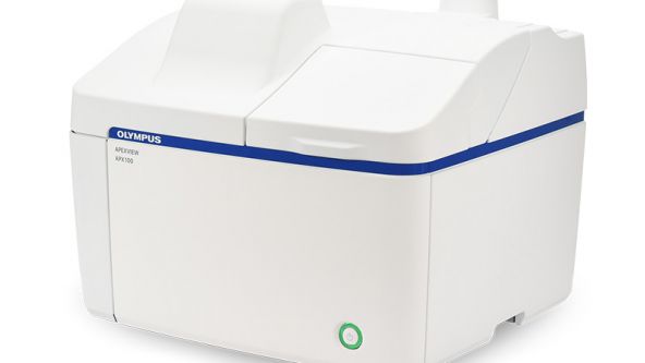
Atsuya Toda is an assistant manager for Global Life Science Marketing at Evident. He has more than fifteen years of experience in life science microscopy sales, sales planning, and marketing in Japan. In 2021, he moved to the Global Life Science Marketing team where he is the marketing representative for the APEXVIEW APX100 all-in-one microscope. He holds a Bachelor of Economics degree from Doshisha University, Japan.
Hikaru Mukai
Since joining the company, Hikaru has been responsible for supporting confocal and super-resolution microscopy products, and she has been a member of the Life Science Marketing department since 2022. Hikaru holds a Bachelor of Science degree from Tokyo University of Science, Japan.

Jake Jones
Jake Jones, PhD is an associate product manager for research imaging at Evident, working with inverted imaging systems, total internal reflection fluorescence (TIRF), fluorescence recovery after photobleaching (FRAP), luminescence, and multiphoton microscopy. As a member of the product management team, Jake works to identify the needs of scientists and researchers and helps provide imaging solutions that match the growing needs of the imaging community. He holds a doctorate in biomedical engineering from the University of Arkansas, where he pursued projects involving biomedical applications of multiphoton microscopy, confocal microscopy, in vivo imaging techniques, and neural network-based image processing.
.jpeg?rev=3653)
Avi Smith
Avi Smith is an associate product manager for research microscopy at Evident. He currently supports the product lines for cell culture, confocal spinning disk, and high-content screening software. Before joining Evident, he spent 10 years working in tissue engineering where he focused on developing skin models for drug discovery and development. Avi holds a master’s degree in engineering management from Tufts University.
Buelent Peker
With over 15 years of experience at Evident, Buelent Peker is a skilled specialist in laser scanning microscopy. His interest in microscopy and photonics began during his doctoral studies in physical chemistry, where he conducted research on time-resolved two-photon microscopy, and his passion for this field has persisted ever since. Buelent has been instrumental in introducing our leading-edge laser scanning microscopes to the market and is particularly intrigued by the potential applications of multiphoton systems as well as the customization options for laser scanning systems.
Kaori Hirayama
Kaori Hirayama目前在Evident的生命科学市场部工作,负责定制产品市场营销。她在拥有10多年的共聚焦显微镜技术支持经验。她拥有日本北里大学医疗卫生学学士学位。

Dr. Peter Su
Michel Biocco
Chloé Savard
Chloé Savard在Instagram上的网名是@tardibabe。她是来自蒙特利尔的微生物学家,三年前开始涉足显微镜的世界。作为一名接受过正式训练的音乐家(她会打鼓),Chloé希望通过学习微生物学来开阔视野,来回答她从小时候就在思考的一些关于存在的问题。在她Instagram的@tardibabe 账号上,Chloé努力将微观世界转化为艺术,同时教育和提高人们对微观生态系统脆弱性的认知。她也喜欢用各种食物和日常用品来做试验。

Makoto Kuwano
Makoto Kuwano在Evident负责显微镜产品开发已有12年的时间。他目前任职于光学开发部,负责物镜的光学设计、组件产品的开发以及产品性能测量技术的开发。他拥有日本东北大学理学硕士学位。

Dr Grace Yuan
Dr. Grace Yuan earned her PhD at the Shanghai Institute of Plant Physiology and Ecology, Chinese Academy of Sciences. She previously worked as a senior scientist in the field of imaging cytometry. Zhenhuan brings her imaging expertise to Evident as an application specialist with a focus on confocal microscopy, multiphoton microscopy, and high-content analysis (HCA).
Shyam Rathod
Shyam Rathod holds a bachelor’s degree in electrical engineering from the Veermata Jijabai Technological Institute (VJTI) in Mumbai, Maharashtra, India. He works as the deputy executive engineer at the Maharashtra State Electricity Transmission Company (MSETCL) under the Government of Maharashtra, India. He is passionate about photomicrography and has been pursuing this unique art form consistently using his own limited resources. His efforts have earned him international recognition, and he is committed to popularizing this unique blend of art and science through consistent and diligent efforts in refining his images.
Amin El-Heliebi
Masatoshi Dehari
After working in sales for biological microscopy in top research fields in Japan, Masatoshi brought his expertise to Evident in 2023. As part of the Life Science Global Marketing team, he is responsible for innovative products such as the APEXVIEW™ APX100 all-in-one microscope and the CM30 incubation monitoring system. He holds a master’s degree from the University of Tokyo.
Kazuhiko Hosono
Kazuhiko Hosono拥有生物物理背景,并获得了早稻田大学理学硕士学位。他于2004年加入Evident,在光学开发和定制解决方案方面拥有超过六年的经验,并在德国汉堡担任过五年的技术支持开发代表。回到日本后,他加入了全球市场部,参与了共聚焦激光扫描显微镜、高性能显微镜物镜和倒置显微镜系统等多种生命科学研究产品的规划、引进和销售拓展工作。

Gianluca Franco
作为Evident意大利生命科学中心的区域经理,Gianluca Franco已有四年多的工作经验,他不仅是一位技术娴熟的专家,而且对光学显微镜和分析技术有着深刻的理解。他对这一领域的兴趣始于大学期间,当时他为了完成自己的生物工程和生物医学工程硕士论文而开始研究荧光显微镜,并一直坚持至今。Gianluca通过演示和介绍先进的成像系统,为显微镜销售团队提供了卓越支持。他通过寻找和开发新的业务机会,为光学显微镜业务的增长提供了支持。

Britta Frenzel
Britta Frenzel has a strong commercial and technical background with a master's in biomedical engineering from Clemson University and several years of experience in the medical device industry. From 2022–2024, Britta supported Evident’s line of benchtop fluorescence microscopes, fixed stage microscopes, and macro zoom fluorescence microscopes on the product management team, where she worked to identify the needs of researchers and provide imaging solutions that match the growing needs of the imaging community.
.jpg?rev=930C)
Akira Saito
Akira Saito studied veterinary medicine at Tokyo University of Agriculture and Technology, Japan, and graduated in 2007. Shortly after, he joined Evident as an application specialist responsible for in vivo imaging systems, high-content analysis systems, and laser confocal systems to support customers in Japan. In 2013, he took over sales promotion for all Evident life science products. In 2018, he moved to Singapore to provide marketing and application support for the APAC market, then moved back to global product marketing in 2023.

Enrico Poege
Enrico Poege is the Global Marketing Communications Lead for Material Science, based in Hamburg, Germany. He holds a diploma in Business Administration from the University of Leipzig and has more than 15 years of experience in marketing and communications.
该作者的文章

Meet the Microscope Performance Monitor: The Ultimate Tool for Core Facility Managers


Collaboration across Borders: Medical Centers Join Forces to Advance Cancer Research

Ask the Experts
Investigating Tumor Dissemination by Spatial Transcriptomics

Transforming Precision Imaging: Meet the FLUOVIEW™ FV4000 Confocal Microscope

Introduction to the APEXVIEW™ APX100 Digital Imaging System

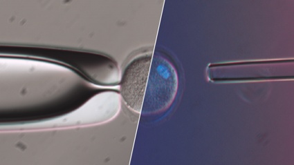
How Polarized Light Can Assist Embryologists in Clinical Routines

Unveiling Nanoscopic Realms: A Journey into Super-Resolution Microscopy
Multiplexing and Deep Tissue Imaging with NIR Confocal Laser Scanning Microscopy (Encore Edition)
Good Cell Culture Practice—How to Improve the Reproducibility of Your Experiments

EVIDENT Organoid Conference 2023 - Looking Deeper, Capturing Complexities
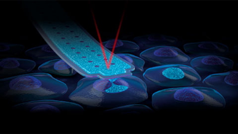
FluidFM:通过直接核内递送进行 CRISPR 基因编辑的新方法
奥林巴斯生物成像会议:探索新维度 | 3天虚拟活动 | 2022年3月9日-11日
Exceptional Imaging Made Easy: Meet the APEXVIEW™ APX100 All-in-One Microscope


Technology Evaluation: Deciphering Cell-Cell Interactions in a 3D Microenvironment at a Single-Cell Resolution

活细胞的超分辨率成像:小物体大格局


FV3000 Red Near-Infrared (NIR) Solutions for Confocal Microscopy | 2 p.m.


FV3000 Red Near-Infrared (NIR) Solutions for Confocal Microscopy | 10 a.m.
Olympus Organoid Conference 2021
.jpg?rev=3E0D)
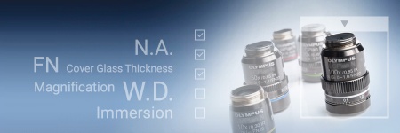
显微镜物镜—魔力之源

高内涵筛查:量身定制的分析让一切变得简单


患者源性类器官和细胞球的三维高通量图像分析

奥林巴斯探索峰会:推进您的成像技术 | 2021 年 10 月 26 日至 27 日
.jpg?rev=1940)
数字图像处理第2讲:高级图像处理之滤波器

对多个亚细胞结构进行纳米级 3D 成像

Whole-Brain Functional Calcium Imaging Using Light Sheet Microscopy

Product Demo: SLIDEVIEW™ VS200 Research Slide Scanner

Product Demo: SLIDEVIEW™ VS200 Research Slide Scanner

The Use of Multiplexing in Microscopy for Better Understanding the Skin Immune System in the Context of the Tissue


Recent Advances in 3D Imaging and AI-Driven Data Analysis

Now You Have the Power to See More

Metabolic Imaging in Langerhans Human Islets with MPE and FLIM

Product Demo: IXplore™ SpinSR Confocal Super Resolution System
In-Vivo Tracking of Harmonic Nanoparticles by Means of a TIGER Widefield Microscope

Hyperspectral and Brightfield Imaging Combined with Deep Learning Uncover Hidden Regularities of Colors and Patterns in Cells and Tissues

Product Demo: FLUOVIEW™ FV3000 Confocal Laser Scanning Microscope
Product Demo: FLUOVIEW™ FV3000 Confocal Laser Scanning Microscope
Evolution of Scientific Digital Imaging Technologies and their Applications

Deep Learning Approaches to Automated Phenotypic Profiling

Deconvolution of 3D Image Stacks
Confocal Microscopy and Its Use for a Spaceflight Experiment

Accelerating Image Analysis with TruAI™ Deep Learning Technology
A New Way of Thinking—Object Detection with Deep Learning
ICSI - How to improve your technique
NoviSight™ Demonstration: 3D Image Analysis and Statistical Software for Organoids and Spheroids
Study the Function of Stromal Cells through Intestinal Organoid Co-Culture Technology

An In Vitro System for Evaluating Anticancer Drugs Using Patient-Derived Tumor Organoids

3D Segmentation for Fluorescence Images: From Qualitative to Quantitative

Prostate Cell Lineage Hierarchy and Plasticity

Investigating Spheroid Architecture Using the FV3000 Confocal Microscope

Advances in 3D Optical Imaging Technologies: An Overview

3D Microscopy: Understanding the Give and Take on Instrument Performance to Enable Informed Decisions

Tissue Optical Clearing Imaging: From In Vitro to In Vivo

Utilizing Tumoroids to Explore Anti-Tumor Immunity in Rectal Cancer

Converting from 2D to 3D: Bio-Techne Solutions for Your 3D Culture

Culture and Quantitative 3D Imaging of Organoids: Challenges and Solutions

A Smarter Approach to Culturing and Nurturing Your Cells


现代全玻片扫描:固定样品的单细胞表型分析


3D 分析:智能软件,深层分析


Modern Slide Scanning: Single-cell Phenotyping on Fixed Samples (Encore Edition)

.jpg?rev=6A21)
To the Diffraction Limit and Beyond: The Nanoscale Organization of Axo-Axonic Synapses | 2 p.m.

To the Diffraction Limit and Beyond: The Nanoscale Organization of Axo-Axonic Synapses | 10 a.m.
Light Sheet Microscopy – New multi-resolution and -color imaging methods

Olympus Organoid Conference: Think Deep, See Deeper | 3-Day Virtual Event | September 7-9, 2021
Create a Smarter Cell Culture Workflow


Digital Image Processing: Point and Local Operation Filters (Encore Edition)
Depth Matters: Transforming Biology for More Realistic and Meaningful Pursuits

.jpg?rev=FD74)
Microscope Objectives—Where the Magic Happens (Encore Edition)
Olympus Discovery Summit—Looking Forward: A New Era of Research
ICSI—Past, Present & Future

Microscope Objectives—Where the Magic Happens


Digital Image Processing: Point and Local Operation Filters

Light Sheet Microscopy for Deeper Insight into Life
Multiplexing and Deep Tissue Imaging with Near-Infrared Confocal Laser Scanning Microscopy
