
Takeo Ogama
Takeo Ogama는 Evident의 선임 제품 및 전략 기획 관리자 겸 현미경 카메라 제품 관리자입니다.Takeo Ogama는 카메라를 비롯하여 다양한 제품을 담당하는 연구 및 개발부에서 8년간 근무했으며 8년간의 기획, 마케팅, 관리 경험을 보유하고 있습니다.그는 일본 오사카대학교에서 중성미자 물리학 석사를 취득했습니다.
Alket Mertiri
Alket Mertiri 박사는 광학 및 포토닉스 분야의 경력을 보유하고 있습니다.Alket Mertiri는 보스턴대학교에서 재료 과학 및 공학 박사를 취득했으며 박사 과정에서 새로운 현미경 기술 개발을 연구했습니다.그는 2018~2019년 Olympus에서 인턴 제품 관리자를 맡아 기본 현미경에서 내부 전반사 현미경 검사와 같은 고급 기술에 이르는 솔루션을 찾음으로써 과학 연구를 발전시켰습니다.
Lauren Alvarenga
Lauren Alvarenga는 Evident의 임상 현미경 부문 선임 제품 관리자입니다.Lauren Alvarenga의 전문 분야는 대물렌즈 및 이미징 소프트웨어입니다.Lauren Alvarenga는 로체스터공과대학교에서 생물 의학 사진 커뮤니케이션 이학사 학위를 취득했습니다.

Ralf Schaefer
Ralf Schaefer는 함부르크대학교에서 화학을 전공했습니다.Ralf Schaefer는 여러 기관 및 회사의 다양한 기술 환경에서 마케팅 커뮤니케이션 부문에 종사했습니다.Ralf Schaefer는 다양한 프로젝트와 역할을 맡으면서 Olympus의 현미경 및 산업 업계의 존재감을 유도 및 형성했습니다.
Kerry Israel
Kerry Israel은 미주 Olympus Corporation의 과학 솔루션 그룹의 생명 과학을 위한 마케팅 및 커뮤니케이션 담당 관리자입니다. Brandeis 대학에서 문학사를 취득하였으며 15년 동안 광고 및 소셜 미디어 전략에서 풀뿌리 홍보에 이르는 모든 측면의 마케팅 경험을 쌓았습니다.

Alec De Grand
Alec De Grand는 생명과학 분야에서 사용되는 Evident의 가상 슬라이드 스캐닝 및 정립 현미경을 담당하는 제품 관리자입니다.Alec De Grand는 Evident에서 10년 이상 근무했으며 근무 기간 동안 임상 제품, 마케팅 기획, 이미징 교육과정 및 박람회를 관리했습니다.
Carlo Alonzo
Carlo Alonzo 박사는 Evident에서 생명과학 현미경 영업 담당자를 맡고 있습니다.Carlo Alonzo는 과학자들이 연구 목표 달성에 도움이 되는 기술을 식별 및 이해하도록 지원합니다.생물 의학 광학에 관심이 있었던 Carlo Alonzo는 보스턴 학계에서 몇 년간 몸담으며 다광자 현미경을 사용하는 연구에 몰두했습니다.Carlo Alonzo는 필리핀대학교에서 물리학 박사 학위를 받았으며 덴마크공과대학교, 하버드 의학전문대학원의 매사추세츠 종합병원에서 박사 후 과정을 밟았습니다.

Joanna Hawryluk
Dr.Joanna Hawryluk는 Evident에서 연구 이미징 제품 관리자로 재직 중입니다.Joanna Hawryluk는 현재 Evident의 세포 배양 배양 모니터링 시스템, 3D 세포 분석 소프트웨어, 전기 생리학 현미경 및 단면광 현미경을 담당하고 있습니다.Joanna Hawryluk는 코네티컷대학교에서 생리학 및 신경 생물학과 박사 학위를 취득했습니다.

Laz Amador
Laz Amador는 Evident에서 국내 응용 팀의 선임 관리자를 맡고 있습니다.Laz Amador는 36년간의 현미경 영업 경험을 보유하고 있으며 1988년부터 Evident에서 근무했습니다.응용 팀에 합류하기 전 Laz Amador는 라틴 아메리카 기술 마케팅 팀의 상임 이사를 맡은 바 있습니다.Laz Amador는 아내 Jan과 플로리다주 웨스턴에서 거주하고 있습니다.
Rebecca Chandler
Rebecca는 Olympus 과학 솔루션의 전속 작가입니다. 엔디콧 대학에서 저널리즘 문학사를 취득하였으며 과학 및 산업의 동향 및 기술에 대해 글을 쓰고 있습니다. Olympus 기술자 및 과학자와 긴밀히 협업하면서 최신 레이저 스캐닝, 초고해상도, 다광자, 정립/실체/도립 현미경 시스템, 첨단 광학, 카메라 및 소프트웨어에 대해 집필하고 있습니다. Olympus의 최신 기사에서 세포학, 병리학, 교육 등 여러 응용 분야에 대한 Rebecca의 글을 확인해 보세요.
Dr. James Lopez
James Lopez는 2010년 시카고대학에서 의생명과학 박사 학위를 취득했습니다.칼슘 이미징, FRET, 생세포 이미징 및 생체 내 이미징 분야에 거의 10년간의 경험이 있는 James는 공초점 및 다광자 솔루션 영업 담당자로 Olympus에 합류했습니다.James는 이후 공초점 및 다광자 시스템을 지원하는 Olympus 생명과학 응용 그룹으로 옮겨갔습니다.이제 그는 미국, 캐나다 및 라틴 아메리카 시장에서 생명과학 응용 그룹을 관리합니다.
.jpg?rev=29C8)
Matthew Weitzman
Matthew Weitzman은 델라웨어대학교에서 생물학 박사 학위를 취득했으며 몇 년에 설친 고급 이미징 경험을 보유하고 있습니다.2014년부터 2021년까지 Matthew Weitzman은 Olympus의 공초점 및 다광자 이미징 시스템을 담당하면서 제품 개발 및 응용 지원을 보조했습니다.
Janeen Manning
Janeen Manning은 Evident에서 임상 및 교육용 현미경의 제품 관리자로 재직 중입니다.Janeen Manning은 Evident에 합류하기 전 감염병 전문 의료 기기 회사에서 근무한 이력이 있습니다.그녀는 메인대학교에서 생화학, 미생물학 및 분자 생물학 학사 학위를, 하버드대학교에서 생명공학 석사 학위를 취득했습니다.
Christopher D. Higgins
Christopher Higgins는 Evident의 생명과학 부문 사업 개발 관리자입니다.Christopher Higgins는 현미경 분야에서 24년간 경험을 쌓은 전문가로, 경력 기간의 대부분을 제품 관리 및 연구 및 임상 응용 분야에 사용되는 다양한 생물 광학 현미경 애플리케이션에 쏟았습니다.
Christopher Higgins는 현재 맡은 직책에서 새로운 기술 개발 및 응용 분야와 관련하여 연구자 및 병리학자들과 직접 협력하고 있습니다.오늘날의 가상 현미경 시스템으로 확보한 이미지들은 개별 세포뿐 아니라 전체 조직에 관한 정보를 제공하며 연구, 교육 등 다양한 분야에 매우 중요합니다.Christopher Higgins는 전체 슬라이드 이미징 및 인공 지능 분석을 준비하여 다음 시대에 진입하는 것이 임상 영역에서 보다 광범위한 응용 분야에 대한 승인을 목표로 하는 상황에서 다음으로 넘어야 할 한계라고 생각합니다.
디지털 병리학 협회 회원인 Christopher Higgins는 마이애미대학교에서 학사 학위를 취득했습니다.Christopher Higgins는 야외 스포츠 애호가이자 뛰어난 자연사진 작가로, 미국 펜실베이니아주 리하이 밸리에 거주 중입니다.
Cheng-Hao Chien
Dr.Cheng-Hao Chien은 대만의 양밍국립대학교에서 생명 광학 박사 학위를 받았으며 보스턴의 터프츠의학대학교의 신경 과학과에서 박사 후 과정을 밟았습니다.고급 현미경 및 생명과학 연구에서 10년간의 경험을 보유한 Cheng-Hao Chien는 2020~2021년 동안 Olympus의 다광자 현미경 및 맞춤형 솔루션을 통해 제품 및 애플리케이션을 지원했습니다.
Brendan Brinkman
Brendan Brinkman은 주요 마우스 신경구 분화 분야에서 몇 년간 근무했습니다.몇 년 동안 핵심 이미징 시설 관리자로 근무한 뒤 Olympus에 합류해 새로운 시스템 구축을 지원했습니다.2017년 Olympus 도쿄 본사에서 차세대 솔루션 개발을 지원하며 2년 반 동안 근무한 뒤 복귀했습니다.
Kathy Lindsley

Manoel Veiga
Manoel Veiga는 물리 화학 박사 학위를 취득했으며 스페인 산티아고데콤포스텔라대학교에서 피코초 및 펨토초 단위의 시간분해 분광법을 연구했습니다.Manoel Veiga는 마드리드 콤플루텐세대학교와 뮌스터대학교에서 박사 후 과정을 두 차례 마친 후 PicoQuant에서 시간분해 분광법, 형광 수명 이미징 현미경(Fluorescence Lifetime Imaging Microscopy, FLIM) 및 형광 상관 분광법(Fluorescence Correlation Spectroscopy, FCS) 분야의 수석 과학자로 근무했습니다.Manoel Veiga는 현재 독일 Evident에서 글로벌 응용 전문가로 재직 중이며 고함량 분석(High-Content Analysis, HCA) 및 딥러닝을 주로 다룹니다.
Heiko Gäthje
Rebecca Bonfig
Dr.Rebecca Bonfig는 루이빌대학교 생리학 및 생물 물리학과에서 대학원 과정을 완료했으며 부갑상선호르몬에 의한 신장 인산염 수송체 Npt2a의 전사 후 조절을 연구했습니다.2015부터 2022년까지, Dr.Rebecca Bonfig는 Olympus의 공초점 현미경 부문에서 근무하면서 제품 관리자로서 FLUOVIEW™ 시리즈를 지원했습니다.
Bülent Peker
Flavio Giacobone
Shohei Imamura
Shohei Imamura는 Evident의 전략 프로젝트 관리자입니다.과학 현미경 영업 부문에서 4년간 근무한 바 있으며 제품 기획, 전략 프로젝트 관리 및 프로젝트 실행 분야에서 7년간의 경험을 보유하고 있습니다.Shohei Imamura는 일본 메이지대학교에서 경영 학사 학위를 취득했습니다.
Daniel Bemmerl
Paul Monnier
Dr. Gulpreet Kaur
Dr.Gulpreet Kaur는 위스콘신매디슨대학교에서 분자 생물학 박사 학위를 취득했으며 생세포 회전 디스크 공초점 현미경을 사용하는 세포 기관 생물학 연구를 진행했습니다.
Dr.Gulpreet Kaur는 현미경 분야에서 6년간의 경험을 보유하고 있으며 공초점, 회전 디스크, 초고분해능, 단면광 이미징 등 다양한 기술 부문에 종사했습니다.Gulpreet Kaur는 위스콘신매디슨대학교의 현미경 및 이미지 분석 핵심 시설에서 교육 담당자로 근무하며 이미징 조건 실험 설계 및 최적화와 관련하여 과학자들과 논의를 진행했습니다.
2018부터 2022년까지 Dr.Gulpreet Kaur는 과학자들이 연구 프로젝트에 Olympus의 현미경 및 이미징 기술을 적용할 수 있도록 지원했습니다.

Dr. Nicolas Bourg
Sarah Williams
2013년에 Olympus의 마케팅 커뮤니케이션부에 입사하기 전에 Sarah는 방송 미디어 산업의 연구원 겸 카피라이터로 약 10년 동안 종사하였습니다. 현재 작가 겸 편집자로서 자신의 스킬을 활용하여 Olympus의 제품 및 전문 기술 분야와 관련된 주제에 관한 고품질 소재를 설득력 있게 다루고 있습니다. 그녀가 집필한 비파괴 검사(NDT) 기술에는 초음파(UT) 및 와전류 탐상 시험(ECT), 페이즈드 어레이(PA), 전체 초점 방법(TFM) 및 풀 매트릭스 캡처(FMC), 산업 현미경 및 비디오내시경이 포함되어 있습니다. 우리를 둘러싼 세계의 품질 및 안전 향상에 있어 Olympus 제품의 기여를 종종 탐구하고 있습니다. 남편인 David와 세 자녀인 Sophie, Anouk, Éloi와 함께 살면서 퀘벡 시 사무실에서 근무하고 있습니다.
Dr. Mike Woerdemann
Mike Woerdemann는 구조형 라이트 필드 기반의 광학 핀셋 기술 분야에서 박사 학위를 취득했습니다.Mike Woerdemann는 몇 년간 고함량 스크리닝 시스템을 지원하는 응용 전문가로 근무했으며, 이 기간 동안 해당 분야에 대한 심도 있는 실용 지식을 확보했습니다.Mike Woerdemann는 현재 고함량 스크리닝 스테이션 및 EVIDENT 유럽 기술센터에서 개발되는 딥러닝 제품의 관리자로 재직 중입니다.

Chunsong Yan
Chunsong Yan은 Olympus 호주 및 뉴질랜드의 생명과학 분야 사업 개발 관리자입니다.그는 현재 공초점, 다광자, 단면광 및 슬라이드 스캐닝 시스템을 담당하고 있습니다.Chunsong Yan은 Evident에 합류한 이후 다양한 역할을 맡으면서 고객에게 최고의 이미징 솔루션을 제공하기 위해 항상 노력하고 있습니다.
Minju Kim
Junsung Kim
Ganesh Kadasoor
Wei Juan Wong
Wei Juan Wong은 Evident의 디지털 슬라이드 스캐닝 시스템의 응용 전문가입니다.Wei Juan Wong은 SLIDEVIEW™ VS200 연구용 슬라이드 스캐너 등 광시야 현미경을 사용하는 동남아시아 고객을 지원하기 위해 싱가포르에서 제품 전문가로 경력을 시작했습니다.이후 Wei Juan Wong은 디지털 슬라이드 스캐닝 시스템 응용 전문가로서 독일에 위치한 EVIDENT 유럽 기술 센터에 합류하여 전 세계 고객들에게 응용 및 마케팅 지원을 제공해 왔습니다.Wei Juan Wong은 물리학 학위를 가지고 있으며 생물 물리학 연구 실험실과 현미경 핵심 시설에서 근무했습니다.
Srivats Hariharan
Stefan Marawske
Bunryu Arashi
Bunryu Arashi는 Evident 유럽 지사의 OEM 관리자로 근무하고 있습니다.12년간의 현미경 제품 개발 경험을 보유한 Bunryu Arashi는 여러 대물렌즈 및 카메라 어댑터뿐만 아니라 BX™ 및 CKX™ 시리즈 현미경용 조명 광학 시스템을 설계했습니다.Bunryu Arashi는 일본 오사카대학교에서 공학 석사 학위를 받았습니다.

Craig Rappaport
Craig Rappaport는 Evident Life Science의 선임 현미경 영업 담당자입니다.Craig Rappaport는 26년의 현미경 영업 경험을 보유하고 있으며 12 년간 과학 및 기술 마케팅 분야에 종사했습니다.Craig Rappaport는 Evident에 합류한 후 시장 분석, 고객 의견 설문 조사 및 신제품 연구를 수행했습니다.2012년부터 Craig Rappaport는 영업 팀에 있으면서 캘리포니아 남부 샌디에이고 및 임페리얼 카운티 지역을 담당했습니다.Craig Rappaport는 캘리포니아대학교 샌타바버라에서 석사 학위를, 듀크대학교 푸쿠아경영대학에서 MBA를 취득했습니다.
Marcus Krauel
Marcus Krauel은 유럽 Evident의 OEM 관리자입니다.그는 12년간 광학 업계에서 쌓은 경험을 바탕으로, 산업용부터 의료용까지 다양한 응용 분야에서 유럽 전역의 Olympus OEM 고객을 지원하고 있습니다.Marcus는 물리 및 광학 엔지니어링 학위를 보유하고 있습니다.
Klaus Willeke
Klaus Willeke는 Evident Life Science에서 제품 마케팅 관리자로 재직 중입니다.그는 22년 이상 Evident에서 근무해 왔습니다.Klaus Willeke는 독일에서 산업 및 생명과학 현미경 영업 담당자로 17년간 근무했으며 이후 유럽 제품 마케팅 팀에 합류하여 정립 임상 및 연구용 현미경과 X Line™ 대물렌즈를 담당하고 있습니다.
Yu Kikuchi
Yu Kikuchi는 Evident의 부품 제품 및 현미경 제품의 광학 엔지니어입니다.Yu Kikuchi는 현미경 제품의 광학 설계 및 평가 경험을 보유하고 있습니다.Yu Kikuchi는 주로 맞춤형 제품 개발, 고객 요구 사항에 대한 사양 맞춤, 현장 테스트 및 조정에 참여했습니다.Yu Kikuchi는 일본 도호쿠대학교에서 분자 생물학 석사를 취득했습니다.
Stephan Fliegel
Stephan Fliegel은 2007년 뮌스터응용과학대학교에서 물리 공학 학사 학위를 취득했으며 레이저 응용 기술을 주로 연구했습니다.Stephan Fliegel은 경력 초기에는 응용 전문가로 근무하다가, 이후 제품 및 프로젝트 관리자를 맡으면서 산업용 레이저 분야에서 고객별 레이저 시스템의 설계 레이아웃, 개발 및 기획을 주로 담당했습니다.현재 Evident에서 광현미경 카메라 제품 관리자로 재직 중입니다.

Dr. Daniel Göttel
Daniel Göttel은 독일 하이델베르크의 독일 암 연구 센터에서 인류 유전학 박사 학위를 취득했습니다.Daniel Göttel은 가상 슬라이드 스캐닝 시스템 응용 전문가로 몇 년간 근무했으며 이 분야에 대한 심도 있는 실용 지식을 보유하고 있습니다.Daniel Göttel은 현재 Evident에서 SLIDEVIEW™ VS200 연구 슬라이드 스캐너 및 Net Image Server(NIS) SQL 영업 지원 관리자를 맡고 있습니다.
Lee Botes
Lee Botes는 Olympus 현미경 영업 경력을 24년간 쌓아왔습니다. 미생물학을 전공한 Lee는 남아프리카 공화국에서 현미경 영업 경력을 시작했습니다.Lee는 뉴질랜드 Olympus 영업 팀의 일원으로써 웰링턴 및 남섬의 산업, 임상 및 연구 현미경과 뉴질랜드의 공초점 현미경 및 슬라이드 스캐너 영업을 담당하고 있습니다.
Masato Yabe
Masato Yabe는 Evident의 광학 개발 부서에서 근무 중이며 19년 동안 현미경 제품 개발에 참여하면서 대물렌즈 및 자동 초점 조절 장치용 광학 시스템을 설계했습니다.Masato Yabe는 일본 와카야마대학교에서 공학 석사를 취득했습니다.
Shodai Hosono
Shodai Hosono는 Evident의 기계 엔지니어링 부서에서 근무하고 있습니다.Shodai Hosono는 10년 이상 현미경 제품 개발에 참여해 왔습니다.Shodai Hosono는 기계 엔지니어로서 다양한 현미경 및 모듈식 현미경 어셈블리를 설계해 왔습니다.Shodai Hosono는 일본 사이타마대학교에서 공학 석사를 취득했습니다.
Kei Sato
Kei Sato는 Evident의 전기 엔지니어링 부서에서 근무 중이며 9년 동안 현미경 제품 개발에 종사해 왔습니다.Kei Sato는 제품 담당자로서 산업 현미경 및 현미경 부품의 전기 설계를 맡고 있습니다.
Joanna Hawryluk
Dr.Joanna Hawryluk는 Evident에서 연구 이미징 제품 관리자로 재직 중입니다.Joanna Hawryluk는 현재 Evident의 세포 배양 배양 모니터링 시스템, 3D 세포 분석 소프트웨어, 전기 생리학 현미경 및 단면광 현미경을 담당하고 있습니다.Joanna Hawryluk는 코네티컷대학교에서 생리학 및 신경 생물학과 박사 학위를 취득했습니다.

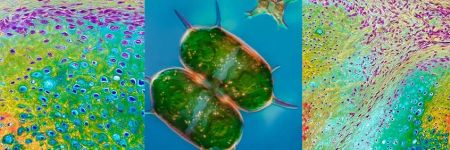
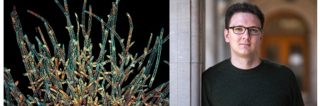
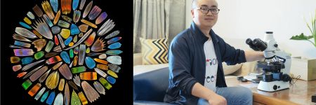
Dr. Hunter Hines
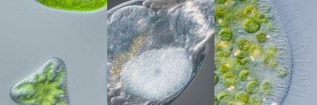
Dr. Hunter Hines received his PhD in 2019 while registered in a joint international collaboration with Bournemouth University (UK) and Harbor Branch Oceanographic Institute (HBOI) at Florida Atlantic University. He conducted research into microbial communities found in tropical aquatic and terrestrial ecosystems in Florida, focusing on the biogeography and biodiversity of ciliates.
Today, Dr. Hines continues his research at HBOI, where he examines microbial communities and the impact they have on ecosystems. He is also the senior scientist on the Amoray Marine Conservation Team at the Amoray SCUBA Resort in Key Largo, Florida.
On social media, Dr. Hines plays a significant science outreach role for microbiology and ecology. During his PhD, he created the Instagram account Microbialecology, which has over 140,000 followers. He is a frequent collaborator with the popular YouTube channel Brave Wilderness, assisting with everything microbial and featuring in several episodes.
Kenji Ono
Kenji Ono는 Olympus의 광학 부품 부서에 재직 중입니다.그는 입사 시점부터 당사의 이미징 사업부에서 디지털카메라의 광학 시스템 설계 및 개발에 참여해 왔습니다.지금은 기존 지식을 광학 분야에 적용하며 OEM 현미경의 마케팅 활동을 총괄하고 있습니다.Kenji Ono는 일본 키타사토대학교에서 학사 학위를 받았습니다.
Kristina Mayer
Kristina Mayer는 Evident 유럽 제품 마케팅 팀의 도립 현미경 제품 마케팅 관리자입니다.Kristina Mayer는 Evident에서 10년 이상 근무했으며 EMEA 지역 내 내부 전반사 형광(Total Internal Reflection Fluorescence, TIRF) 현미경 및 회전 디스크 공초점 현미경을 포함한 연구 이미징을 지원하는 IXplore™ 도립 현미경 시리즈를 담당하고 있습니다.Kristina Mayer는 야콥스대학교 브레멘에서 세포 생물학 박사 학위를 취득했습니다.
Benjamin Compans, Ph.D.
Yuan Yue
Yuan Yue는 Evident의 시장 전략 부서 소속으로, 주로 디지털 마케팅을 맡고 있습니다.Yuan Yue는 Evident에서 6년에 가깝게 마케팅 커뮤니케이션 업무를 담당했습니다.
Shogo Usui
Dr. Anne Beghin
Dr. Xiaotong Cui
Dr. Kasmira Wilson
Dr. Dan Zhu
Dr. Graham Wright
Mr. Srivats Hariharan

Ms. Gency Gunasingh
Dr. Dong Gao

Dr. Yu Weimiao
Dr. Motoki Takagi
Dr. Ningbo Wu
.jpg?rev=C8C3)
Mr. Hiroya Ishihara
Marie Kawasaki
Marie Kawasaki는 Evident의 생명과학 마케팅 부서 소속입니다.Marie Kawasaki는 7년 동안 임상 연구와 관련된 현미경 마케팅에 종사했으며 현재 불임 치료 연구(세포질 내 정자 주입) 산업을 대상으로 글로벌 영업 프로모션을 담당하고 있습니다.
Wei Juan Wong
Wei Juan Wong은 Evident의 디지털 슬라이드 스캐닝 시스템의 응용 전문가입니다.Wei Juan Wong은 SLIDEVIEW™ VS200 연구용 슬라이드 스캐너 등 광시야 현미경을 사용하는 동남아시아 고객을 지원하기 위해 싱가포르에서 제품 전문가로 경력을 시작했습니다.이후 Wei Juan Wong은 디지털 슬라이드 스캐닝 시스템 응용 전문가로서 독일에 위치한 EVIDENT 유럽 기술 센터에 합류하여 전 세계 고객들에게 응용 및 마케팅 지원을 제공해 왔습니다.Wei Juan Wong은 물리학 학위를 가지고 있으며 생물 물리학 연구 실험실과 현미경 핵심 시설에서 근무했습니다.
Samuel M Lawrence
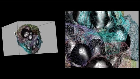
Samuel M. Lawrence is the chief executive officer and cofounder of CytoViva, Inc. He led the development of the core, patented optical illumination technology that is the foundation of CytoViva’s products, as well as played a key role in the development of the patented CytoViva 3D imaging technology. He continues to lead all product development efforts and actively supports the company’s sales efforts both domestically and internationally.
Manoel Veiga
Manoel Veiga는 물리 화학 박사 학위를 취득했으며 스페인 산티아고데콤포스텔라대학교에서 피코초 및 펨토초 단위의 시간분해 분광법을 연구했습니다.Manoel Veiga는 마드리드 콤플루텐세대학교와 뮌스터대학교에서 박사 후 과정을 두 차례 마친 후 PicoQuant에서 시간분해 분광법, 형광 수명 이미징 현미경(Fluorescence Lifetime Imaging Microscopy, FLIM) 및 형광 상관 분광법(Fluorescence Correlation Spectroscopy, FCS) 분야의 수석 과학자로 근무했습니다.Manoel Veiga는 현재 독일 Evident에서 글로벌 응용 전문가로 재직 중이며 고함량 분석(High-Content Analysis, HCA) 및 딥러닝을 주로 다룹니다.
Dr. Manfred Kässens
Dr.Manfred Kässens는 EVIDENT 유럽 기술센터에서 기술 제품 정보 부서를 이끌고 있으며 Evident의 글로벌 마케팅 커뮤니케이션 부서 팀원으로 재직 중입니다.Manfred Kässens는 독일 뮌스터대학교에서 물리학 박사를 취득했으며 1997년부터 Evident에서 근무하고 있습니다.
Sara Quiñones Gonzalez
Sara Quiñones González는 생명 공학 학위를 보유했습니다.EVIDENT 유럽 기술센터에 제품 관리자로 합류하기 전에는, 여러 연구실 및 임상 실험실에서 근무했습니다.Sara Quiñones González는 VS200 연구용 슬라이드 스캐너를 포함한 SLIDEVIEW 스캐너 시리즈 업무를 담당하고 있습니다.
Dr. Thomas Bauer-Jazayeri

Maria Ada Prusicki
Maria Ada Prusicki는 2019년에 함부르크대학교에서 생물학 박사 학위를 취득했습니다.Maria Ada Prusicki는 박사 과정 동안 식물 세포 분열을 추적하기 위한 생세포 이미징 기술을 집중 연구했습니다.박사 후 과정 중에는 현미경 기술에 대한 지식을 계속해서 심화했습니다.2022년, Maria Ada Prusicki는 SLIDEVIEW™ VS200 연구용 슬라이드 스캐너의 응용 전문가가 되었습니다.Evident의 독일 뮌스터 사무소에서 근무 중인 Maria Ada Prusicki는 Evident의 글로벌 영업 및 마케팅 팀뿐 아니라 전 세계 고객에게 VS200 응용 지원을 제공하고 있습니다.
Taichi Yoshikura
Taichi Yoshikura는 일본 도쿄 Evident에서 글로벌 생명과학 마케팅 커뮤니케이션 팀을 이끌고 있습니다.Taichi Yoshikura는 와세다대학교에서 문학사 학위를 받았으며 2016년부터 Evident에 재직 중입니다.
Takuma Saito
Takuma Saito는 일본 도쿄 Evident 산업 현미경 영업 팀에서 2011년부터 근무했습니다.Takuma Saito는 일본의 전자 부품 제조업체들과 강력한 관계를 구축한 후 현재 당사의 레이저 스캐닝 현미경 및 디지털 현미경 제품의 영업 개발 프로젝트를 이끌고 있습니다.
Dr. Yongjie Wang
Dr. Yongjie Wang은중국 베이징에 있는 Evident Life Science의 응용 전문가입니다.그녀는 현재 고급 현미경의 과학적 응용에 중점을 두고 있습니다.Dr.Wang은 난징대학교에서 화학 및 화학 공학 박사 학위를 취득했습니다.

Koji Nakagawa
Koji Nakagawa는 12년간 디지털카메라 개발에 종사했으며 이미징 렌즈의 광학 시스템 설계를 감독했습니다.현재는 Evident의 광학 개발 부서에서 현미경 제품을 개발하고 있습니다.Koji Nakagawa는 일본 야마나시대학교에서 공학 석사를 취득했습니다.
Atsuya Toda
Atsuya Toda is an assistant manager for Global Life Science Marketing at Evident. He has more than fifteen years of experience in life science microscopy sales, sales planning, and marketing in Japan. In 2021, he moved to the Global Life Science Marketing team where he is the marketing representative for the APEXVIEW APX100 all-in-one microscope. He holds a Bachelor of Economics degree from Doshisha University, Japan.
Hikaru Mukai
Since joining the company, Hikaru has been responsible for supporting confocal and super-resolution microscopy products, and she has been a member of the Life Science Marketing department since 2022. Hikaru holds a Bachelor of Science degree from Tokyo University of Science, Japan.

Jake Jones
Jake Jones, PhD is an associate product manager for research imaging at Evident, working with inverted imaging systems, total internal reflection fluorescence (TIRF), fluorescence recovery after photobleaching (FRAP), luminescence, and multiphoton microscopy. As a member of the product management team, Jake works to identify the needs of scientists and researchers and helps provide imaging solutions that match the growing needs of the imaging community. He holds a doctorate in biomedical engineering from the University of Arkansas, where he pursued projects involving biomedical applications of multiphoton microscopy, confocal microscopy, in vivo imaging techniques, and neural network-based image processing.
.jpeg?rev=3653)
Avi Smith
Avi Smith is an associate product manager for research microscopy at Evident. He currently supports the product lines for cell culture, confocal spinning disk, and high-content screening software. Before joining Evident, he spent 10 years working in tissue engineering where he focused on developing skin models for drug discovery and development. Avi holds a master’s degree in engineering management from Tufts University.
Buelent Peker
With over 15 years of experience at Evident, Buelent Peker is a skilled specialist in laser scanning microscopy. His interest in microscopy and photonics began during his doctoral studies in physical chemistry, where he conducted research on time-resolved two-photon microscopy, and his passion for this field has persisted ever since. Buelent has been instrumental in introducing our leading-edge laser scanning microscopes to the market and is particularly intrigued by the potential applications of multiphoton systems as well as the customization options for laser scanning systems.
Kaori Hirayama
Kaori Hirayama는 현재 Evident의 생명과학 마케팅 부서에서 맞춤형 제품 마케팅을 담당하고 있습니다. 컨포컬 현미경 지원 분야에서 10년 이상의 경험을 축적했으며, 일본 키타사토 대학교(Kitasato University)에서 위생 기술 학사 학위를 취득했습니다.
Dr. Peter Su
Michel Biocco
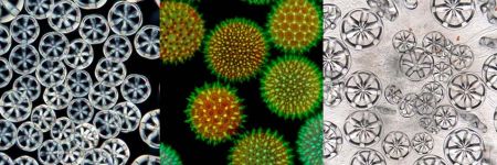
Chloé Savard
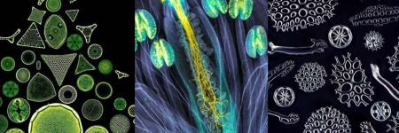
인스타그램에서 @tardibabe로 활동하는 Chloé Savard는 3년 전 현미경의 세계에 대해서 알게 된 몬트리올 출신의 미생물학자입니다. 드러머로써 뮤지션의 길을 걷던 Chloe는 견문을 넓히고 어린 시절부터 가진 존재론적 질문에 답하기 위해 미생물학을 공부하기로 했습니다. @tardibabe 인스타그램 채널을 통해 현미경 생태계의 취약성을 알리고 이에 대한 인식을 높이는 동시에 인식할 수 없는 대상을 예술로 승화하기 위해 노력하고 있습니다. 그녀는 다양한 음식과 일상적 물건들로 실험하는 것을 좋아합니다.

Makoto Kuwano
Makoto Kuwano는 Evident에서 12년 동안 현미경 제품 개발 업무를 담당해 왔습니다. 그는 현재 광학 개발 사업부에서 대물렌즈 광학 설계, 부품 개발, 제품 성능을 위한 측정 기법 개발에 참여하고 있습니다. Makoto는 일본 도호쿠 대학교에서 이학 석사를 취득했습니다.

Dr Grace Yuan
Dr. Grace Yuan earned her PhD at the Shanghai Institute of Plant Physiology and Ecology, Chinese Academy of Sciences. She previously worked as a senior scientist in the field of imaging cytometry. Zhenhuan brings her imaging expertise to Evident as an application specialist with a focus on confocal microscopy, multiphoton microscopy, and high-content analysis (HCA).
Shyam Rathod
Shyam Rathod holds a bachelor’s degree in electrical engineering from the Veermata Jijabai Technological Institute (VJTI) in Mumbai, Maharashtra, India. He works as the deputy executive engineer at the Maharashtra State Electricity Transmission Company (MSETCL) under the Government of Maharashtra, India. He is passionate about photomicrography and has been pursuing this unique art form consistently using his own limited resources. His efforts have earned him international recognition, and he is committed to popularizing this unique blend of art and science through consistent and diligent efforts in refining his images.
Amin El-Heliebi
Masatoshi Dehari
After working in sales for biological microscopy in top research fields in Japan, Masatoshi brought his expertise to Evident in 2023. As part of the Life Science Global Marketing team, he is responsible for innovative products such as the APEXVIEW™ APX100 all-in-one microscope and the CM30 incubation monitoring system. He holds a master’s degree from the University of Tokyo.
Kazuhiko Hosono
Kazuhiko Hosono는 생물물리학 분야에서 경력을 쌓아 왔으며 와세다대학에서 이학석사를 취득했습니다. 2004년에 Evident에 입사한 후 광학 개발 및 맞춤형 솔루션 분야에서 6년 넘게 경력을 쌓은 다음 5년간 독일 함부르크에서 개발 담당자로서 기술 지원을 제공했습니다. 일본으로 돌아온 후에는 글로벌 마케팅 부서에 합류하여 컨포컬 레이저 스캐닝 현미경, 고성능 현미경 대물렌즈, 도립 현미경 시스템 등 광범위한 생명 과학 연구 제품의 기획, 도입, 영업 확장에 관여했습니다.

Gianluca Franco
As territory manager for Evident’s Life Science Center, Italy, for over four years, Gianluca Franco is a skilled specialist with a strong understanding of optical microscopes and analysis techniques. His interest in this field began during university while working on fluorescence microscopy for his master’s thesis in bioengineering and biomedical engineering, and it has persisted ever since. Gianluca provides outstanding support to the team for microscope sales by delivering demonstrations and presentations of advanced imaging systems. He provides support for the growth of the optical microscopy business by identifying and developing new business opportunities.

Britta Frenzel
Britta Frenzel has a strong commercial and technical background with a master's in biomedical engineering from Clemson University and several years of experience in the medical device industry. From 2022–2024, Britta supported Evident’s line of benchtop fluorescence microscopes, fixed stage microscopes, and macro zoom fluorescence microscopes on the product management team, where she worked to identify the needs of researchers and provide imaging solutions that match the growing needs of the imaging community.
.jpg?rev=930C)
Akira Saito
Akira Saito studied veterinary medicine at Tokyo University of Agriculture and Technology, Japan, and graduated in 2007. Shortly after, he joined Evident as an application specialist responsible for in vivo imaging systems, high-content analysis systems, and laser confocal systems to support customers in Japan. In 2013, he took over sales promotion for all Evident life science products. In 2018, he moved to Singapore to provide marketing and application support for the APAC market, then moved back to global product marketing in 2023.

Enrico Poege
Enrico Poege is the Global Marketing Communications Lead for Material Science, based in Hamburg, Germany. He holds a diploma in Business Administration from the University of Leipzig and has more than 15 years of experience in marketing and communications.
Posts by this author


Ask the Experts
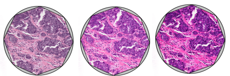
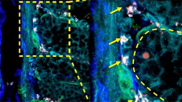
Investigating Tumor Dissemination by Spatial Transcriptomics

Transforming Precision Imaging: Meet the FLUOVIEW™ FV4000 Confocal Microscope

Introduction to the APEXVIEW™ APX100 Digital Imaging System

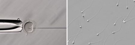
How Polarized Light Can Assist Embryologists in Clinical Routines

Unveiling Nanoscopic Realms: A Journey into Super-Resolution Microscopy
Multiplexing and Deep Tissue Imaging with NIR Confocal Laser Scanning Microscopy (Encore Edition)
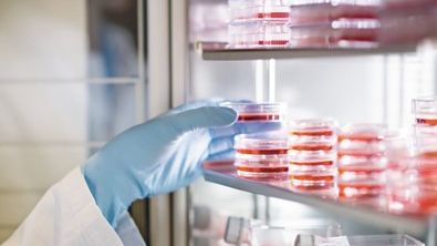
Good Cell Culture Practice—How to Improve the Reproducibility of Your Experiments

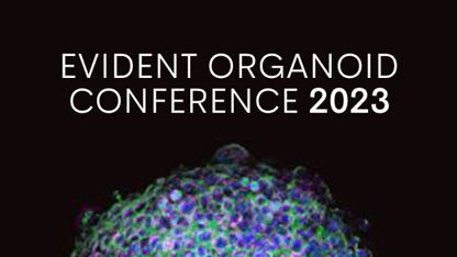
EVIDENT Organoid Conference 2023 - Looking Deeper, Capturing Complexities
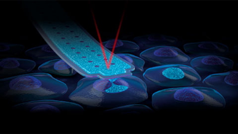
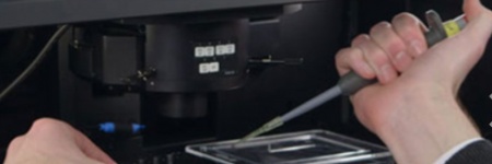
FluidFM: 핵 내 직접 전달을 통한 새로운 CRISPR 유전자 편집 방식
Exceptional Imaging Made Easy: Meet the APEXVIEW™ APX100 All-in-One Microscope


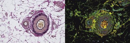
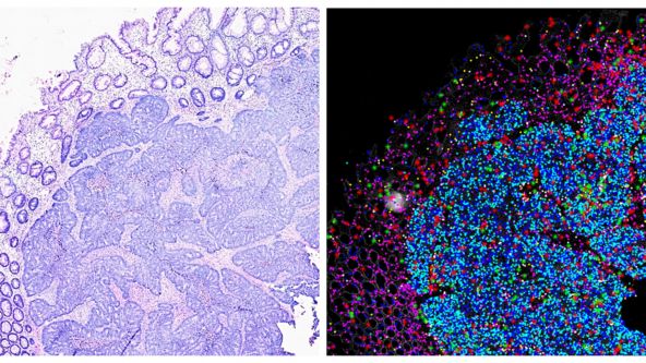
최신 슬라이드 스캐닝: 고정 샘플에서의 단세포 표현형


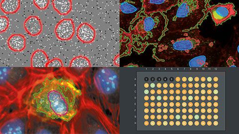
Technology Evaluation: Deciphering Cell-Cell Interactions in a 3D Microenvironment at a Single-Cell Resolution

3D 분석: 지능형 소프트웨어, 통찰력 있는 분석


딥러닝: 새 애플리케이션의 문을 열다


Olympus 바이오이미징 콘퍼런스: 새로운 차원 탐구 | 3일간의 가상 이벤트 | 2022년 3월 9~11일
라이브 셀 초고해상도 이미징: 작은 대상을 큰 그림으로


FV3000 Red Near-Infrared (NIR) Solutions for Confocal Microscopy | 2 p.m.


FV3000 Red Near-Infrared (NIR) Solutions for Confocal Microscopy | 10 a.m.
Olympus Organoid Conference 2021
.jpg?rev=3E0D)
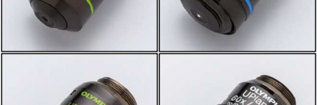
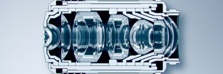
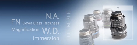
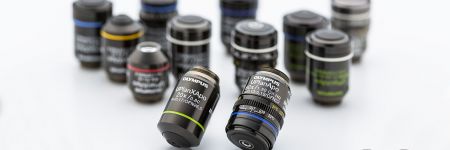
현미경 대물렌즈 - 마법이 펼쳐지는 곳

High-Content 스크리닝: 간편해진 맞춤형 분석
나노미터 크기의 여러 세포 내 구조를 3D로 이미징하기

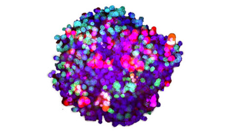
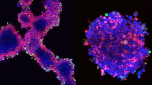
환자 유래 오가노이드 및 스페로이드의 3차원 High-Throughput 이미지 분석

Olympus 디스커버리 서밋: 이미징 향상 | 2021년 10월 26일~27일
.jpg?rev=1940)
디지털 이미지 처리 2부: 고급 이미지 처리 필터

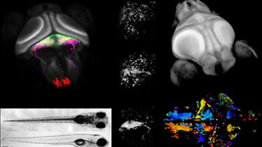
Whole-Brain Functional Calcium Imaging Using Light Sheet Microscopy

Product Demo: SLIDEVIEW™ VS200 Research Slide Scanner

Product Demo: SLIDEVIEW™ VS200 Research Slide Scanner

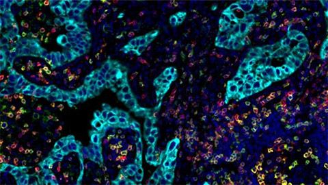
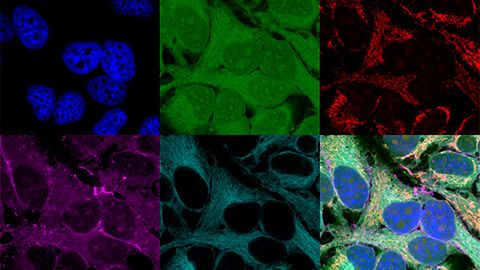
The Use of Multiplexing in Microscopy for Better Understanding the Skin Immune System in the Context of the Tissue


Recent Advances in 3D Imaging and AI-Driven Data Analysis

Now You Have the Power to See More

Metabolic Imaging in Langerhans Human Islets with MPE and FLIM

Product Demo: IXplore™ SpinSR Confocal Super Resolution System
In-Vivo Tracking of Harmonic Nanoparticles by Means of a TIGER Widefield Microscope

Hyperspectral and Brightfield Imaging Combined with Deep Learning Uncover Hidden Regularities of Colors and Patterns in Cells and Tissues

Product Demo: FLUOVIEW™ FV3000 Confocal Laser Scanning Microscope
Product Demo: FLUOVIEW™ FV3000 Confocal Laser Scanning Microscope
Evolution of Scientific Digital Imaging Technologies and their Applications


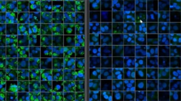
Deep Learning Approaches to Automated Phenotypic Profiling

Deconvolution of 3D Image Stacks
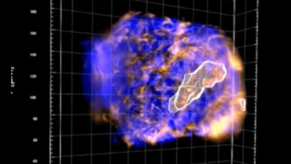
Confocal Microscopy and Its Use for a Spaceflight Experiment

Accelerating Image Analysis with TruAI™ Deep Learning Technology
A New Way of Thinking—Object Detection with Deep Learning
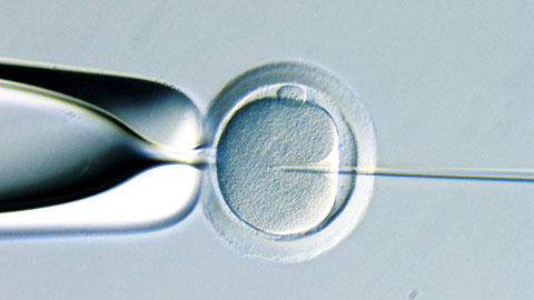
ICSI - How to improve your technique
NoviSight™ Demonstration: 3D Image Analysis and Statistical Software for Organoids and Spheroids
Study the Function of Stromal Cells through Intestinal Organoid Co-Culture Technology

An In Vitro System for Evaluating Anticancer Drugs Using Patient-Derived Tumor Organoids

3D Segmentation for Fluorescence Images: From Qualitative to Quantitative

Prostate Cell Lineage Hierarchy and Plasticity

Investigating Spheroid Architecture Using the FV3000 Confocal Microscope

Advances in 3D Optical Imaging Technologies: An Overview

3D Microscopy: Understanding the Give and Take on Instrument Performance to Enable Informed Decisions

Tissue Optical Clearing Imaging: From In Vitro to In Vivo

Utilizing Tumoroids to Explore Anti-Tumor Immunity in Rectal Cancer

Converting from 2D to 3D: Bio-Techne Solutions for Your 3D Culture

Culture and Quantitative 3D Imaging of Organoids: Challenges and Solutions

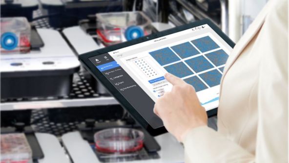
A Smarter Approach to Culturing and Nurturing Your Cells

Modern Slide Scanning: Single-cell Phenotyping on Fixed Samples (Encore Edition)

.jpg?rev=6A21)
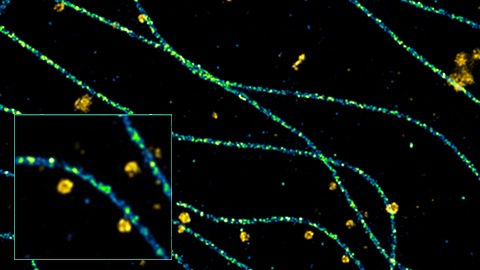
To the Diffraction Limit and Beyond: The Nanoscale Organization of Axo-Axonic Synapses | 2 p.m.

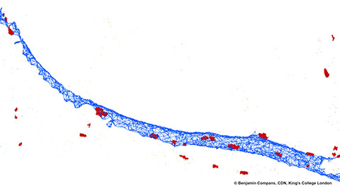
To the Diffraction Limit and Beyond: The Nanoscale Organization of Axo-Axonic Synapses | 10 a.m.
Light Sheet Microscopy – New multi-resolution and -color imaging methods

Olympus Organoid Conference: Think Deep, See Deeper | 3-Day Virtual Event | September 7-9, 2021
Create a Smarter Cell Culture Workflow


Digital Image Processing: Point and Local Operation Filters (Encore Edition)
Depth Matters: Transforming Biology for More Realistic and Meaningful Pursuits

.jpg?rev=FD74)
Microscope Objectives—Where the Magic Happens (Encore Edition)
Olympus Discovery Summit—Looking Forward: A New Era of Research
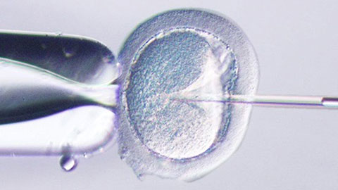
ICSI—Past, Present & Future

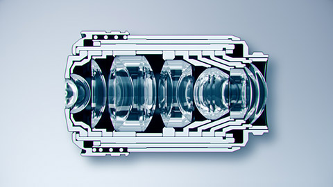
Microscope Objectives—Where the Magic Happens

Think Deep, See Deeper with Near-Infrared Laser Scanning Confocal Microscope

Digital Image Processing: Point and Local Operation Filters

Light Sheet Microscopy for Deeper Insight into Life
Multiplexing and Deep Tissue Imaging with Near-Infrared Confocal Laser Scanning Microscopy
