
小蒲 健夫
小蒲健夫氏はEvidentのシニアプロダクト・戦略プランナーであり、顕微鏡カメラのプロダクトマネージャーも務めています。カメラを含むさまざまな製品の研究・開発部門で8年、製品計画、マーケティング、マネジメントで8年の経験を有しています。大阪大学でニュートリノ物理学の修士号を取得。
Alket Mertiri
Alket Mertiri氏(PhD)は光学および光工学のバックグラウンドがあります。ボストン大学で材料科学および工学の博士号を取得し、大学では最新の検鏡法技術の開発に携わっていました。2018年~2019年にオリンパスでアソシエイトプロダクトマネージャーを務め、基本的な検鏡法から全反射照明検鏡法のような高度な技術に至るソリューションを模索し、科学者たちの研究を進歩させました。
Lauren Alvarenga
Lauren Alvarenga氏は、Evidentで臨床用顕微鏡のシニアプロダクトマネージャーを務めています。専門に担当しているのは対物レンズとイメージングソフトウェアです。ロチェスター工科大学で生物医学フォトグラフィックコミュニケーションの理学士号を取得しています。

Ralf Schaefer
Ralf Schaefer氏はハンブルク大学で化学の学位を取得しています。いくつかの代理店や企業で、さまざまな技術環境でのマーケティングコミュニケーションを担当しました。Evidentでは、各種プロジェクトや担当業務で、検鏡法の認知度と産業ビジネスを加速させ具体化させてきました。
Kerry Israel
Kerry Israel氏は、Olympus Corporation of the AmericasのScientific Solutions Groupのライフサイエンスのマーケティングおよびコミュニケーションのマネージャーです。Brandeis Universityで文学士号を取得し、広告・ソーシャルメディア戦略からグラスルーツアウトリーチまで、マーケティングのすべての側面で15年を超える経験を有しています。

Alec De Grand
Alec De Grand氏は、Evidentのライフサイエンス用バーチャルスライドスキャンおよび正立顕微鏡のプロダクトマネージャーです。10年以上Evidentに勤務し、その間、臨床製品、マーケティングイニシアチブ、イメージングコース、展示会などに携わりました。
Carlo Alonzo
Carlo Alonzo氏(PhD)はEvidentでライフサイエンス顕微鏡の営業担当を務めています。科学者たちが研究目標を達成するための技術を特定し、理解できるように支援を行っています。生物医学光学系に興味を持ち、ボストンの学会に参加するようになってから、多光子検鏡法を用いた研究に数年間没頭しました。フィリピン大学で物理学の博士号を取得し、デンマーク工科大学とマサチューセッツ総合病院(ハーバード・メディカルスクール)で博士課程修了後のトレーニングを積みました。

Joanna Hawryluk
Dr. Joanna Hawrylukは、Evidentのリサーチイメージングのプロダクトマネージャーです。現在は、細胞培養インキュベーションモニタリングシステム、3D細胞解析ソフトウェア、電気生理学顕微鏡、ライトシート顕微鏡を担当しています。コネチカット大学の生理学・神経生物学部で博士号を取得しています。

Laz Amador
Laz Amador氏は、Evidentナショナルアプリケーションチームのシニアマネージャーです。顕微鏡のセールスで36年の経験を持ち、1988年からEvidentに在籍しています。アプリケーションチームに加わる前は、ラテンアメリカ地域のテクニカルマーケティング執行役員でした。妻のJanさんとともにフロリダ州ウエストン在住。
Rebecca Chandler
Rebecca氏は、Olympus Scientific Solutionsのスタッフライターです。Endicott Collegeでジャーナリズムの学士号を取得し、科学および産業におけるトレンドとテクノロジーについて執筆しています。オリンパスのエンジニアや科学者と密接につながって仕事をし、最新のレーザー走査型、超解像、多光子、正立型、実体、倒立型の顕微鏡システムの他、最先端の光学系、カメラ、ソフトウェアについての記事を書いています。細胞学、病理学、教育など、数多くのアプリケーションにおけるオリンパスの最新の状況を知るためには、彼女の記事を読んでください。

Dr. James Lopez
James Lopez氏は、2010年にシカゴ大学で生物医科学の博士号を取得しました。カルシウムイメージング、FRET、ライブセルイメージング、生体イメージングの10年近い経験を活かし、共焦点・多光子顕微鏡のセールス担当者としてEvidentに入社しました。後に、共焦点・多光子システムをサポートするEvidentライフサイエンスアプリケーショングループに異動しました。現在、米国、カナダ、中南米の市場で、ライフサイエンスアプリケーショングループを管理しています。
.jpg?rev=29C8)
Matthew Weitzman
Matthew Weitzman氏はデラウェア大学で生物科学の博士号を取得し、高度なイメージングに関する長年の経験を有しています。2014年~2021年、オリンパスの共焦点・多光子イメージングシステムに取り組み、製品開発やアプリケーションサポートを支援しました。
Janeen Manning
Janeen Manning氏は、Evidentの臨床・教育用顕微鏡のプロダクトマネージャーです。Evident入社前は、感染症専門の医療機器会社に勤務していました。メイン大学で生物化学、微生物学、分子生物学の理学士号を取得し、ハーバード大学でバイオテクノロジーの修士号(MLA)を取得しています。
Christopher D. Higgins
Christopher Higgins氏は、Evidentライフサイエンス部門の事業開発マネージャーです。24年に及ぶ検鏡法分野での経験があり、研究と臨床の両アプリケーションで用いられる広範な生物学的光学顕微鏡のプロダクトマネジメントとアプリケーションでキャリアを積み重ねてきました。
現在の役職では、研究者や病理学者と直接協同して新たな技術とアプリケーションの開発に取り組んでいます。今日のバーチャル顕微鏡システムで撮影した画像は、個別の細胞だけでなく組織全体についてもわかることがあり、研究や教育などに不可欠なものになっています。Higgins氏は、次の大きな開拓は、臨床分野でのより広範なアプリケーションの受け入れに向かう次の時代に入るためのホールスライドイメージングと人工知能による解析を準備することだと考えます。
同氏はDigital Pathology Associationのメンバーであり、マイアミ大学で学士号を取得しています。また、熱心な野外活動家でもあり、自然写真家として成功し、ペンシルベニア州のリーハイ・バレーに住んでいます。
Cheng-Hao Chien
Dr. Cheng-Hao Chienは、台湾の国立陽明大学でバイオフォトニクスの博士号を取得し、ボストンのタフツ大学医学部神経科学科で博士課程修了後のトレーニングを積みました。高度な検鏡法とライフサイエンス研究における10年間の経験を活かし、2020年~2021年にオリンパスで多光子検鏡法とカスタマイズソリューションを担当し、製品とアプリケーションのサポートを担いました。
Brendan Brinkman
Brendan Brinkman氏は、何年もの間、マウスニューロスフェアの一次分離を含む幹細胞分野で研究を続けてきました。コアイメージング施設のマネージャーとして数年間勤務し、新システム構築の支援を行った後にオリンパスに入社しました。オリンパス東京本社に2年半勤務した後、2017年に米国に戻りました。東京では、次世代ソリューションの開発を担っていました。
Kathy Lindsley

Manoel Veiga
Manoel Veiga氏は、サンティアゴ・デ・コンポステーラ大学(スペイン)でピコ秒およびフェムト秒時間分解分光法を研究し、物理化学の博士号を取得しました。マドリード・コンプルテンセ大学とミュンスター大学で2つのポスドク研究を行った後、PicoQuant社でシニアサイエンティストとして勤務し、時間分解分光法、蛍光寿命イメージング観察法(FLIM)、蛍光相関分光法(FCS)の分野で研究しました。現在はドイツのEvidentでグローバルアプリケーションスペシャリストとして、ハイコンテント解析(HCA)とディープラーニングを中心に業務に携わっています。
Heiko Gäthje
Rebecca Bonfig
Dr. Rebecca Bonfigは、ルイビル大学の生理学・生物物理学部で、副甲状腺ホルモンによる腎リン酸輸送体Npt2aの転写後調節について研究し、大学院を修了しました。2015年~2022年はオリンパスの共焦点顕微鏡を担当し、プロダクトマネージャーとしてFLUOVIEW™ シリーズをサポートしました。
Bülent Peker
Flavio Giacobone
今村 尚平
今村尚平氏は、Evidentの戦略プロジェクトマネージャーを務めています。ライフサイエンス向け顕微鏡のセールスで4年、製品計画、戦略的プロジェクトマネジメント、プロジェクト実行において7年の経験を積んでいます。明治大学で商学士号を取得。
Daniel Bemmerl
Paul Monnier
Dr. Gulpreet Kaur
Dr. Gulpreet Kaurは、ウィスコンシン大学マディソン校で、生細胞のスピニングディスク共焦点検鏡法を用いたオルガネラ生物学を研究し、分子生物学の博士課程を修了しました。
Dr. Kaurは、6年間の検鏡法の経験を持ち、共焦点、スピニングディスク、超解像、ライトシートイメージングなどの様々な技術を手掛けてきました。ウィスコンシン大学マディソン校の検鏡法/画像解析コア施設のトレーナーを務め、実験デザインとイメージング条件の最適化について科学者たちと意見を交換してきました。
2018年~2022年は、科学者がオリンパスの顕微鏡およびイメージング技術を研究プロジェクトに応用する際の支援を行いました。

Dr. Nicolas Bourg
Sarah Williams
Sarah Williams氏は、2013年にオリンパスのマーケティングコミュニケーション部に入る前、放送メディア産業で調査員兼コピーライターとして10年近く働いてきました。現在は、ライター、編集者としてのスキルを活かし、オリンパスの製品や専門技術に関連するトピックについて興味深く高品質な資料を作成しています。彼女が非破壊試験(nondestructive testing:NDT)技術について書いたものでは、超音波(UT)と渦電流試験(eddy current testing:ECT)、フェーズドアレイ(PA)、トータルフォーカシングメソッド(TFM)とフルマトリックキャプチャ(FMC)、および工業用顕微鏡とビデオスコープについて取り上げています。私達を取り巻く世界の品質と安全性の向上に対するオリンパス製品の貢献についても折りに触れ詳しく検証しています。ケベックシティのオフィスに勤務し、同じケベックシティ内に夫のDavidと3人の子供、Sophie、Anouk、Éloiと一緒に暮らしています。
Dr. Mike Woerdemann
Mike Woerdemann氏は、構造化ライトフィールドに基づく光ピンセット技術の博士号を取得しました。アプリケーションスペシャリストとして勤務し、ハイコンテントスクリーニングシステムを長年にわたりサポートする中で、この分野での実践的な知見を深めました。現在は、EVIDENT Technology Center Europeで開発されたハイコンテントスクリーニングステーションとディープラーニング製品のプロダクトマネージャーを務めています。
Chunsong Yan
Chunsong Yan氏は、オーストラリアとニュージーランドのEvidentでライフサイエンス事業開発マネージャーを務めています。現在、共焦点、多光子励起、ライトシート、およびスライドスキャンシステムを担当しています。Evidentに入社して以来、常に最高のイメージングソリューションをお客様に提案しながら、様々な役割を担当してきました。
Minju Kim
Junsung Kim
Ganesh Kadasoor
Wei Juan Wong
Wei Juan Wong氏は、Evidentでデジタルスライドスキャンシステムのアプリケーションスペシャリストを務めています。シンガポールのプロダクトスペシャリストとして当社に加わり、東南アジア地域で、SLIDEVIEW™ VS200リサーチスライドスキャナーなどの広視野顕微鏡を使用するお客様をサポートしました。その後ドイツに移り、EVIDENT Technology Center Europeにアプリケーションスペシャリストとして勤務し、世界中のお客様にアプリケーションとマーケティングのサポートを提供しています。物理学の学位を取得し、生物物理学研究室と顕微鏡法コア施設に勤務した経験があります。
Srivats Hariharan
Stefan Marawske
嵐文隆
嵐文隆氏は欧州EvidentでOEMマネージャーを務めています。12年間にわたる顕微鏡製品の開発経験を持ち、対物レンズやカメラアダプター、BX™ およびCKX™ シリーズ顕微鏡の照明光学系設計を担当してきました。大阪大学で工学修士号を取得。

Craig Rappaport
Craig Rappaport氏は、Evident Life Scienceの顕微鏡のシニアセールス担当者です。26年に及ぶ顕微鏡の営業経験に加え、科学/技術分野のマーケティングにおいても10年余りの経験があります。Evidentに入社以来、市場分析、顧客の声の調査、新製品のリサーチを専門としています。2012年から、セールスチームとともに南カリフォルニアのサンディエゴとインペリアル郡を担当しています。カリフォルニア大学サンタバーバラ校で学士号(BA)、デューク大学のFuqua School of BusinessでMBAを取得しています。
Marcus Krauel
Marcus Krauel氏は欧州EvidentでOEMマネージャーを務めています。12年にわたる光学系を扱ってきた経験を基に、欧州全域のOEMのお客様を、工業分野から医療分野まで幅広いアプリケーションにわたり支援しています。物理学と光工学の学位を取得しています。
Klaus Willeke
Klaus Willeke氏はEvident Life Scienceのプロダクトマーケティングマネージャーです。Evidentに22年あまり勤務しています。ドイツで17年間、工業用・ライフサイエンス用顕微鏡のセールス担当として勤務した後、European Product Marketingチームで、正立型臨床用、研究用顕微鏡およびX Line™ 対物レンズを担当しています。
菊地 由宇
Evidentで装置組み込み用コンポーネント製品、顕微鏡製品の光学エンジニアを務めています。顕微鏡製品の光学設計・評価の実績があり、カスタマイズ製品の開発や顧客要件に合わせた仕様のマッチング、現場でのテストやアライメントに取り組んでいます。東北大学で分子生物学の修士号を取得。
Stephan Fliegel
Stephan Fliegel氏は、ミュンスター応用科学大学でレーザーアプリケーション技術の研究に取り組み、2007年に物理工学の学位を取得しました。工業用レーザー製品の分野で顧客固有のレーザーシステムの設計レイアウト、開発、プランニングを中心に、アプリケーションスペシャリストとして勤務した後、製品およびプロジェクトのマネージャーを務めました。現在は、Evidentで光学顕微鏡用カメラのプロダクトマネージャーを務めています。

Dr. Daniel Göttel
Daniel Göttel氏は、ハイデルベルク(ドイツ)にあるドイツがん研究センターで人類遺伝学の博士号を取得しました。バーチャルスライドスキャニングシステムのアプリケーションスペシャリストとして長年勤務し、この分野で深く実践的な経験を積んできました。現在はEvidentで、SLIDEVIEW™ VS200リサーチスライドスキャナーおよびネットイメージサーバー(NIS)SQLのセールスサポートマネージャーを務めています。
Lee Botes
Lee Botes氏は、24年にわたりオリンパス顕微鏡のセールスを経験してきました。微生物学のバックグラウンドを活かし、南アフリカで顕微鏡セールスのキャリアをスタートさせました。ニュージーランドのEvidentセールスチームの一員として、ウェリントンおよびサウスアイランドで工業用、医療用、研究用の顕微鏡を担当するほかに、ニュージーランド全土で共焦点顕微鏡とスライドスキャナーも担当しています。
矢部 正人
Evident光学開発部門に所属し、19年間にわたって顕微鏡製品の開発に携わり、対物レンズやオートフォーカスの光学系設計を担当しています。和歌山大学で工学修士号を取得。
細野 翔大
Evidentメカニカルエンジニアリング部門に所属。10年以上にわたり顕微鏡製品の開発に携わっています。メカニカルエンジニアとして、各種顕微鏡、コンポーネント顕微鏡を設計しました。埼玉大学で工学士号を取得。
佐藤 慶
Evidentの電気開発部門に所属し、9年間にわたり顕微鏡製品の開発に携わっています。プロダクトリーダーとして、工業用顕微鏡やコンポーネント顕微鏡の電気設計を担当しています。
Joanna Hawryluk
Dr. Joanna Hawrylukは、Evidentのリサーチイメージングのプロダクトマネージャーです。現在は、細胞培養インキュベーションモニタリングシステム、3D細胞解析ソフトウェア、電気生理学顕微鏡、ライトシート顕微鏡を担当しています。コネチカット大学の生理学・神経生物学部で博士号を取得しています。

Dr. Hunter Hines
Dr. Hunter Hines received his PhD in 2019 while registered in a joint international collaboration with Bournemouth University (UK) and Harbor Branch Oceanographic Institute (HBOI) at Florida Atlantic University. He conducted research into microbial communities found in tropical aquatic and terrestrial ecosystems in Florida, focusing on the biogeography and biodiversity of ciliates.
Today, Dr. Hines continues his research at HBOI, where he examines microbial communities and the impact they have on ecosystems. He is also the senior scientist on the Amoray Marine Conservation Team at the Amoray SCUBA Resort in Key Largo, Florida.
On social media, Dr. Hines plays a significant science outreach role for microbiology and ecology. During his PhD, he created the Instagram account Microbialecology, which has over 140,000 followers. He is a frequent collaborator with the popular YouTube channel Brave Wilderness, assisting with everything microbial and featuring in several episodes.
小野 憲司
Evidentのオプティカルコンポーネント部門に所属。入社以来、映像事業でデジタルカメラの光学系の開発・企画に携わってきました。光学知識を活かし、現在はOEM顕微鏡のマーケティング活動に取り組んでいます。北里大学で学士号取得。
Kristina Mayer
Kristina Mayer氏は、EvidentのEuropean Product Marketingチームで倒立顕微鏡のプロダクトマーケティングマネージャーを務めています。10年以上にわたりEvidentに勤務し、IXplore™ 倒立顕微鏡シリーズを担当して、EMEA地域でリサーチイメージング(全反射照明検鏡法(TIRF)やスピニングディスク型共焦点顕微鏡など)をサポートしています。ヤーコプス大学ブレーメンで細胞生物学の博士号を取得しています。
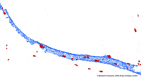
Benjamin Compans, Ph.D.
Yuan Yue
Evidentの市場戦略部門で、主にデジタルマーケティングを担当しています。Evidentのマーケティングコミュニケーション部門で6年近くの経験を有しています。
Shogo Usui
Dr. Anne Beghin
Dr. Xiaotong Cui
Dr. Kasmira Wilson
Dr. Dan Zhu
Dr. Graham Wright
Mr. Srivats Hariharan
Ms. Gency Gunasingh
Dr. Dong Gao

Dr. Yu Weimiao
Dr. Motoki Takagi
Dr. Ningbo Wu
.jpg?rev=C8C3)
Mr. Hiroya Ishihara
川崎 眞莉栄
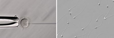
Evidentライフサイエンスマーケティング部門に所属。臨床研究に関する顕微鏡のマーケティングに7年間携わり、現在は不妊治療研究(卵細胞質内精子注入法)産業のグローバルセールスプロモーションを担当しています。
Wei Juan Wong
Wei Juan Wong氏は、Evidentでデジタルスライドスキャンシステムのアプリケーションスペシャリストを務めています。シンガポールのプロダクトスペシャリストとして当社に加わり、東南アジア地域で、SLIDEVIEW™ VS200リサーチスライドスキャナーなどの広視野顕微鏡を使用するお客様をサポートしました。その後ドイツに移り、EVIDENT Technology Center Europeにアプリケーションスペシャリストとして勤務し、世界中のお客様にアプリケーションとマーケティングのサポートを提供しています。物理学の学位を取得し、生物物理学研究室と顕微鏡法コア施設に勤務した経験があります。
Samuel M Lawrence
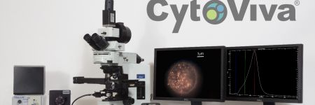
Samuel M. Lawrence is the chief executive officer and cofounder of CytoViva, Inc. He led the development of the core, patented optical illumination technology that is the foundation of CytoViva’s products, as well as played a key role in the development of the patented CytoViva 3D imaging technology. He continues to lead all product development efforts and actively supports the company’s sales efforts both domestically and internationally.
Manoel Veiga
Manoel Veiga氏は、サンティアゴ・デ・コンポステーラ大学(スペイン)でピコ秒およびフェムト秒時間分解分光法を研究し、物理化学の博士号を取得しました。マドリード・コンプルテンセ大学とミュンスター大学で2つのポスドク研究を行った後、PicoQuant社でシニアサイエンティストとして勤務し、時間分解分光法、蛍光寿命イメージング観察法(FLIM)、蛍光相関分光法(FCS)の分野で研究しました。現在はドイツのEvidentでグローバルアプリケーションスペシャリストとして、ハイコンテント解析(HCA)とディープラーニングを中心に業務に携わっています。
Dr. Manfred Kässens
Dr. Manfred Kässensは、EVIDENT Technology Center Europeのテクニカル製品情報部門を率いているとともに、Evidentグローバルマーケティングコミュニケーション部門の一員でもあります。ドイツのミュンスター大学で物理学の修士号を取得し、1997年以来Evidentに勤務しています。
Sara Quiñones Gonzalez
Sara Quiñones González氏はバイオテクノロジーの学位を取得しています。いくつかの研究・臨床検査機関で勤務した後、プロダクトマネージャーとしてEVIDENT Technology Center Europeに加わりました。現在、VS200リサーチスライドスキャナーなどのSLIDEVIEW™ スキャナーシリーズを担当しています。
Dr. Thomas Bauer-Jazayeri

Maria Ada Prusicki
Maria Ada Prusicki氏は、2019年にハンブルク大学で生物学の博士号を取得しています。博士課程では、植物細胞分裂を追跡するライブセルイメージング法に重点をおいて研究していました。ポスドク研究を進める中で、顕微鏡法の知識を深めていきました。2022年に当社SLIDEVIEW™ VS200リサーチスライドスキャナーのアプリケーションスペシャリストになりました。Evidentミュンスターオフィス(ドイツ)を拠点として、世界中のお客様とEvidentのグローバルセールスおよびマーケティングチームに向けて、VS200アプリケーションサポートを提供しています。
吉倉 太一
Evident東京オフィスのグローバルライフサイエンス・マーケティングコミュニケーションチームに所属。早稲田大学で学士号を取得し、2016年からEvidentに勤務しています。
斉藤 拓真
2011年から、Evident東京オフィスの工業用顕微鏡セールスチームの一員です。日本の電子部品メーカーと強力な関係を構築し、現在はレーザー走査型顕微鏡とデジタルマイクロスコープ製品のセールス開発プロジェクトを指揮しています。
Dr. Yongjie Wang
Dr. Yongjie Wangは、北京(中国)のEvident Life Science拠点で活躍するアプリケーションスペシャリストです。現在は、先進的な顕微鏡の科学的応用に注力しています。Dr. Wangは南京大学化学化工学院で博士号を取得しました。

中川 孝司
12年間にわたってデジタルカメラの開発に携わり、撮像レンズの光学系設計を担当しました。現在はEvidentの光学開発部門に所属し、顕微鏡製品の開発に取り組んでいます。山梨大学で工学修士号を取得。
戸田敦哉
日本市場において生物顕微鏡の営業担当、販売企画業務を15年以上にわたり経験したのち、2021年よりライフサイエンスグローバルマーケティングチームにてAPEXVIEW APX100の製品担当を務めています。
向井ひかる
入社以来共焦点顕微鏡や超解像顕微鏡製品のサポートを担った後、2022年からライフサイエンスリサーチマーケティング部に所属。日本の東京理科大学で学士(理学)号取得。

Jake Jones
Jake Jones, PhD is an associate product manager for research imaging at Evident, working with inverted imaging systems, total internal reflection fluorescence (TIRF), fluorescence recovery after photobleaching (FRAP), luminescence, and multiphoton microscopy. As a member of the product management team, Jake works to identify the needs of scientists and researchers and helps provide imaging solutions that match the growing needs of the imaging community. He holds a doctorate in biomedical engineering from the University of Arkansas, where he pursued projects involving biomedical applications of multiphoton microscopy, confocal microscopy, in vivo imaging techniques, and neural network-based image processing.
.jpeg?rev=3653)
Avi Smith
Avi Smith is an associate product manager for research microscopy at Evident. He currently supports the product lines for cell culture, confocal spinning disk, and high-content screening software. Before joining Evident, he spent 10 years working in tissue engineering where he focused on developing skin models for drug discovery and development. Avi holds a master’s degree in engineering management from Tufts University.
Buelent Peker
With over 15 years of experience at Evident, Buelent Peker is a skilled specialist in laser scanning microscopy. His interest in microscopy and photonics began during his doctoral studies in physical chemistry, where he conducted research on time-resolved two-photon microscopy, and his passion for this field has persisted ever since. Buelent has been instrumental in introducing our leading-edge laser scanning microscopes to the market and is particularly intrigued by the potential applications of multiphoton systems as well as the customization options for laser scanning systems.
Kaori Hirayama
Kaori Hirayama currently works in the Life Science Marketing department at Evident where she is in charge of marketing customize products. She has over 10 years of experience working in confocal microscopy support. She holds a Bachelor of Hygienic Technology degree from Kitasato University, Japan.
Dr. Peter Su
Michel Biocco
Chloé Savard
Chloé Savardさんは、Instagramでは@tardibabeとしても知られるモントリオールの微生物学者です。音楽家(ドラマー)として教育を受けた後、視野を広げて子供の頃から抱いていたいくつかの疑問を解き明かすべく、微生物学を学ぶ決心をしました。彼女の@tardibabe Instagramチャンネルでは、微小なものをアートに変えることを目指すとともに、微細なエコシステムの脆弱性について情報を提供し、関心を集めています。また、さまざまな食べ物や日用品を使った実験も大好きです。
桑野信
2011年にオリンパス株式会社に入社し、12年間にわたって顕微鏡製品の開発を担当。現在は株式会社エビデント光学開発部門に所属し、対物レンズの光学設計やコンポーネント製品の開発、製品性能の測定技術開発などに取り組む。東北大学で理学修士号を取得。

Dr Grace Yuan
Dr. Grace Yuan earned her PhD at the Shanghai Institute of Plant Physiology and Ecology, Chinese Academy of Sciences. She previously worked as a senior scientist in the field of imaging cytometry. Zhenhuan brings her imaging expertise to Evident as an application specialist with a focus on confocal microscopy, multiphoton microscopy, and high-content analysis (HCA).
Shyam Rathod
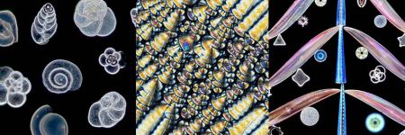
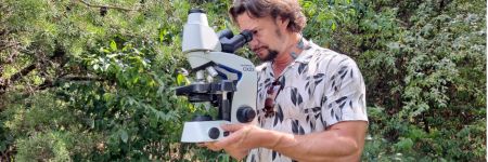
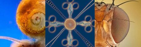
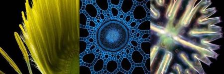
Shyam Rathod holds a bachelor’s degree in electrical engineering from the Veermata Jijabai Technological Institute (VJTI) in Mumbai, Maharashtra, India. He works as the deputy executive engineer at the Maharashtra State Electricity Transmission Company (MSETCL) under the Government of Maharashtra, India. He is passionate about photomicrography and has been pursuing this unique art form consistently using his own limited resources. His efforts have earned him international recognition, and he is committed to popularizing this unique blend of art and science through consistent and diligent efforts in refining his images.
Amin El-Heliebi
Masatoshi Dehari
After working in sales for biological microscopy in top research fields in Japan, Masatoshi brought his expertise to Evident in 2023. As part of the Life Science Global Marketing team, he is responsible for innovative products such as the APEXVIEW™ APX100 all-in-one microscope and the CM30 incubation monitoring system. He holds a master’s degree from the University of Tokyo.
Kazuhiko Hosono
生物物理学のバックグラウンドがあり、早稲田大学で理学修士を取得。2004年にEvidentに入社し、光学開発とカスタマイズソリューション部門を6年以上経験した後、ハンブルグ(ドイツ)で開発担当としてテクニカルサポートを5年経験。日本に戻ってからはグローバルマーケティング部門に加わり、共焦点レーザー走査型、高性能対物レンズ、倒立顕微鏡システムなど、各種ライフサイエンスリサーチ製品の計画、導入、販売拡大に取り組んでいます。

Gianluca Franco
Evident Life Science Center(イタリア)のテリトリーマネージャーを約4年務め、光学顕微鏡と分析法に関して深い見識を持つスペシャリスト。この分野への興味は、生体工学と医用生体工学の修士論文のために蛍光観察について研究したことをきっかけに、それ以来続いている。高度なイメージングシステムのデモンストレーションとプレゼンテーションを担当し、顕微鏡の販売に貢献している。新たなビジネスチャンスを見極め、広げることで、光学顕微鏡事業の成長を支えている。

Britta Frenzel
Britta Frenzel has a strong commercial and technical background with a master's in biomedical engineering from Clemson University and several years of experience in the medical device industry. From 2022–2024, Britta supported Evident’s line of benchtop fluorescence microscopes, fixed stage microscopes, and macro zoom fluorescence microscopes on the product management team, where she worked to identify the needs of researchers and provide imaging solutions that match the growing needs of the imaging community.
.jpg?rev=930C)
Akira Saito
Akira Saito studied veterinary medicine at Tokyo University of Agriculture and Technology, Japan, and graduated in 2007. Shortly after, he joined Evident as an application specialist responsible for in vivo imaging systems, high-content analysis systems, and laser confocal systems to support customers in Japan. In 2013, he took over sales promotion for all Evident life science products. In 2018, he moved to Singapore to provide marketing and application support for the APAC market, then moved back to global product marketing in 2023.

Enrico Poege
Enrico Poege is the Global Marketing Communications Lead for Material Science, based in Hamburg, Germany. He holds a diploma in Business Administration from the University of Leipzig and has more than 15 years of experience in marketing and communications.
この著者による投稿


Ask the Experts
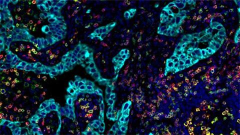
Investigating Tumor Dissemination by Spatial Transcriptomics

Transforming Precision Imaging: Meet the FLUOVIEW™ FV4000 Confocal Microscope

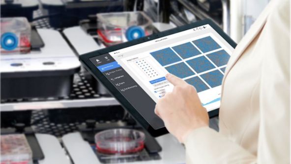
Introduction to the APEXVIEW™ APX100 Digital Imaging System

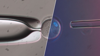
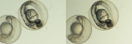
How Polarized Light Can Assist Embryologists in Clinical Routines

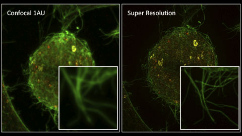
Unveiling Nanoscopic Realms: A Journey into Super-Resolution Microscopy
Multiplexing and Deep Tissue Imaging with NIR Confocal Laser Scanning Microscopy (Encore Edition)
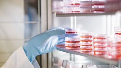
Good Cell Culture Practice—How to Improve the Reproducibility of Your Experiments

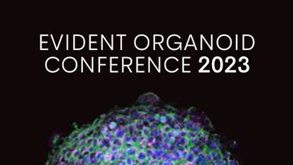
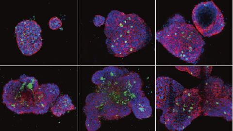
EVIDENT Organoid Conference 2023 - Looking Deeper, Capturing Complexities
FluidFM:核内直接送達によるCRISPR遺伝子編集への新たな手法
Exceptional Imaging Made Easy: Meet the APEXVIEW™ APX100 All-in-One Microscope


最新のスライドスキャン:固定サンプルにおけるシングルセル表現型解析


Technology Evaluation: Deciphering Cell-Cell Interactions in a 3D Microenvironment at a Single-Cell Resolution

3Dアッセイ:高性能なソフトウェアで洞察力に優れた分析


ディープラーニング:新たなアプリケーションへの扉を開く


オリンパス・バイオイメージング・カンファレンス:新次元の探索 | 3日間のバーチャルイベント | 2022年3月9日~11日
ライブセル超解像イメージング: 小さなものの全体像


FV3000 Red Near-Infrared (NIR) Solutions for Confocal Microscopy | 2 p.m.


FV3000 Red Near-Infrared (NIR) Solutions for Confocal Microscopy | 10 a.m.
オリンパス・ディスカバリー・サミット:イメージング力を高めよう | 2021年10月26~27日
Olympus Organoid Conference 2021
.jpg?rev=3E0D)
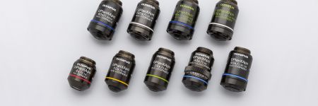
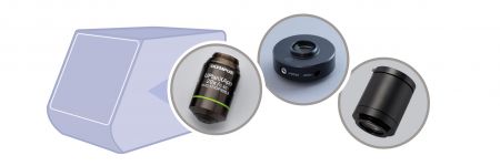
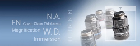
顕微鏡対物レンズ:魔法が起こる場所

ハイコンテントスクリーニング:カスタム解析を簡単に
患者由来のオルガノイドとスフェロイドの三次元高スループット画像解析

.jpg?rev=1940)
デジタル画像処理パート2:先進の画像処理フィルター

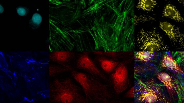
複数の細胞内構造体に対するナノスケールの3Dイメージング

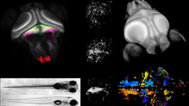
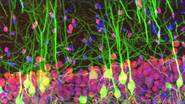
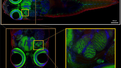
Whole-Brain Functional Calcium Imaging Using Light Sheet Microscopy

Product Demo: SLIDEVIEW™ VS200 Research Slide Scanner

Product Demo: SLIDEVIEW™ VS200 Research Slide Scanner

The Use of Multiplexing in Microscopy for Better Understanding the Skin Immune System in the Context of the Tissue


Recent Advances in 3D Imaging and AI-Driven Data Analysis

Now You Have the Power to See More

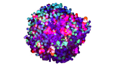
Metabolic Imaging in Langerhans Human Islets with MPE and FLIM

Product Demo: IXplore™ SpinSR Confocal Super Resolution System
In-Vivo Tracking of Harmonic Nanoparticles by Means of a TIGER Widefield Microscope

Hyperspectral and Brightfield Imaging Combined with Deep Learning Uncover Hidden Regularities of Colors and Patterns in Cells and Tissues

Product Demo: FLUOVIEW™ FV3000 Confocal Laser Scanning Microscope
Product Demo: FLUOVIEW™ FV3000 Confocal Laser Scanning Microscope
Evolution of Scientific Digital Imaging Technologies and their Applications

Deep Learning Approaches to Automated Phenotypic Profiling

Deconvolution of 3D Image Stacks
Confocal Microscopy and Its Use for a Spaceflight Experiment

Accelerating Image Analysis with TruAI™ Deep Learning Technology
A New Way of Thinking—Object Detection with Deep Learning
ICSI - How to improve your technique
NoviSight™ Demonstration: 3D Image Analysis and Statistical Software for Organoids and Spheroids
Study the Function of Stromal Cells through Intestinal Organoid Co-Culture Technology

An In Vitro System for Evaluating Anticancer Drugs Using Patient-Derived Tumor Organoids

3D Segmentation for Fluorescence Images: From Qualitative to Quantitative

Prostate Cell Lineage Hierarchy and Plasticity

Investigating Spheroid Architecture Using the FV3000 Confocal Microscope

Advances in 3D Optical Imaging Technologies: An Overview

3D Microscopy: Understanding the Give and Take on Instrument Performance to Enable Informed Decisions

Tissue Optical Clearing Imaging: From In Vitro to In Vivo

Utilizing Tumoroids to Explore Anti-Tumor Immunity in Rectal Cancer

Converting from 2D to 3D: Bio-Techne Solutions for Your 3D Culture

Culture and Quantitative 3D Imaging of Organoids: Challenges and Solutions

A Smarter Approach to Culturing and Nurturing Your Cells

Modern Slide Scanning: Single-cell Phenotyping on Fixed Samples (Encore Edition)

.jpg?rev=6A21)
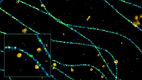
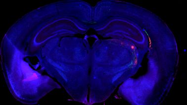
To the Diffraction Limit and Beyond: The Nanoscale Organization of Axo-Axonic Synapses | 2 p.m.

To the Diffraction Limit and Beyond: The Nanoscale Organization of Axo-Axonic Synapses | 10 a.m.
Light Sheet Microscopy – New multi-resolution and -color imaging methods

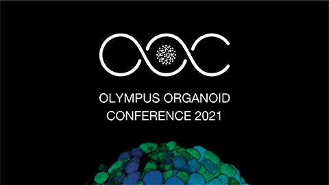
Olympus Organoid Conference: Think Deep, See Deeper | 3-Day Virtual Event | September 7-9, 2021
Create a Smarter Cell Culture Workflow


Digital Image Processing: Point and Local Operation Filters (Encore Edition)
Depth Matters: Transforming Biology for More Realistic and Meaningful Pursuits

.jpg?rev=FD74)
Microscope Objectives—Where the Magic Happens (Encore Edition)
Olympus Discovery Summit—Looking Forward: A New Era of Research
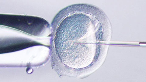
ICSI—Past, Present & Future

Microscope Objectives—Where the Magic Happens

Think Deep, See Deeper with Near-Infrared Laser Scanning Confocal Microscope

Digital Image Processing: Point and Local Operation Filters

Light Sheet Microscopy for Deeper Insight into Life
Multiplexing and Deep Tissue Imaging with Near-Infrared Confocal Laser Scanning Microscopy
