Olympus Image of the Year Award 2019Der Image of the Year Award 2019 ist zu Ende gegangen. Schauen Sie sich im Folgenden die wunderschönen Bilder an, die dieses Jahr eingereicht wurden. Alle aktuellen Informationen und Teilnahmebedingungen erfahren Sie hier! |
|
Globaler Gewinner
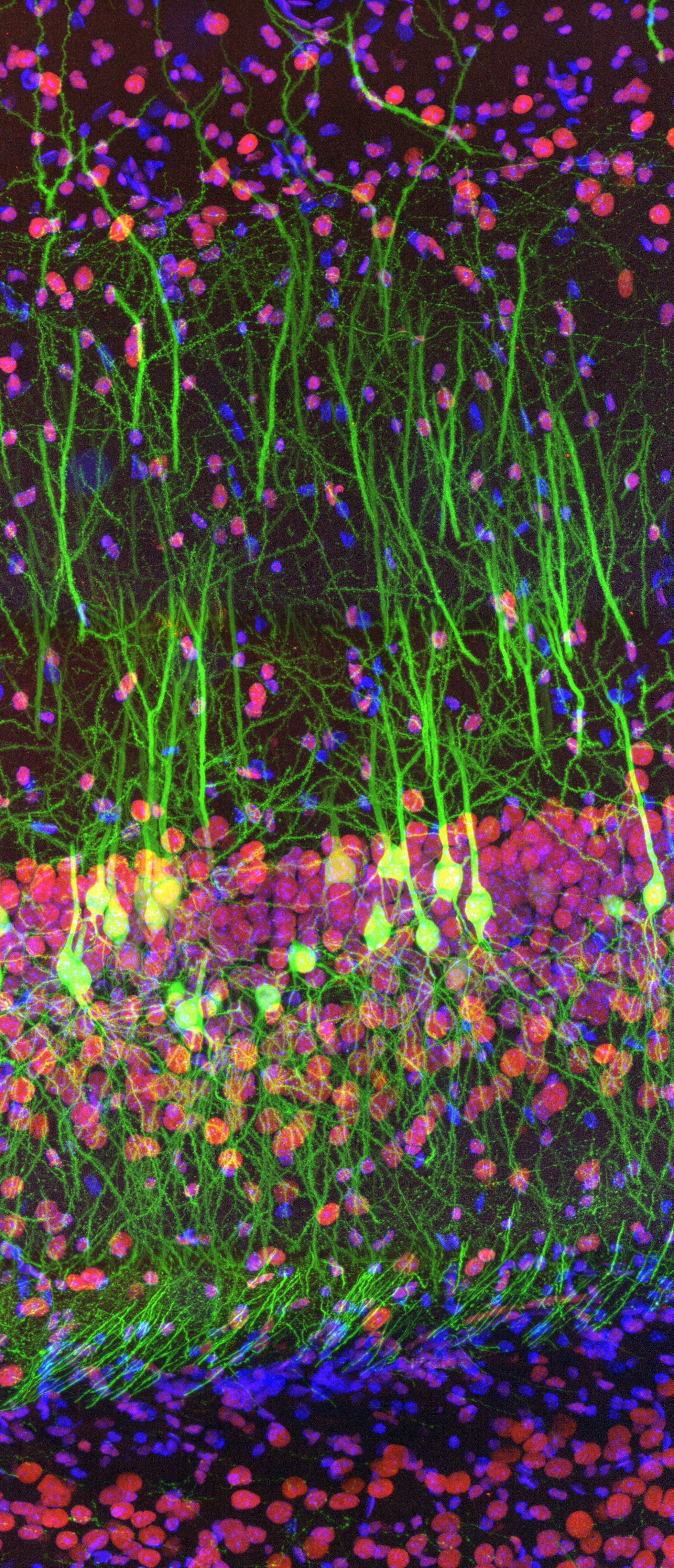 | Das Siegerbild wurde von Ainara Pintor (Spanien) aufgenommen. Das beeindruckende Fluoreszenzbild zeigt den mit zwei Fluorophoren immungefärbten Schnitt eines Mausgehirns. Grün: Exzitatorische Neuronen des Hippocampus, die grün fluoreszierendes Protein unter dem Thy1 -Promotor exprimieren. Rot: Das Fettmasse- und Adipositas-assoziierte Protein (FTO) wurde mit dem Antikörper Alexa Fluor 594 markiert. Blau: Mit DAPI markierte Zellkerne. Aufgenommen mit einem Mikroskopsystem mit Super-Resolution-Technologie |
Regionale Gewinner
Americas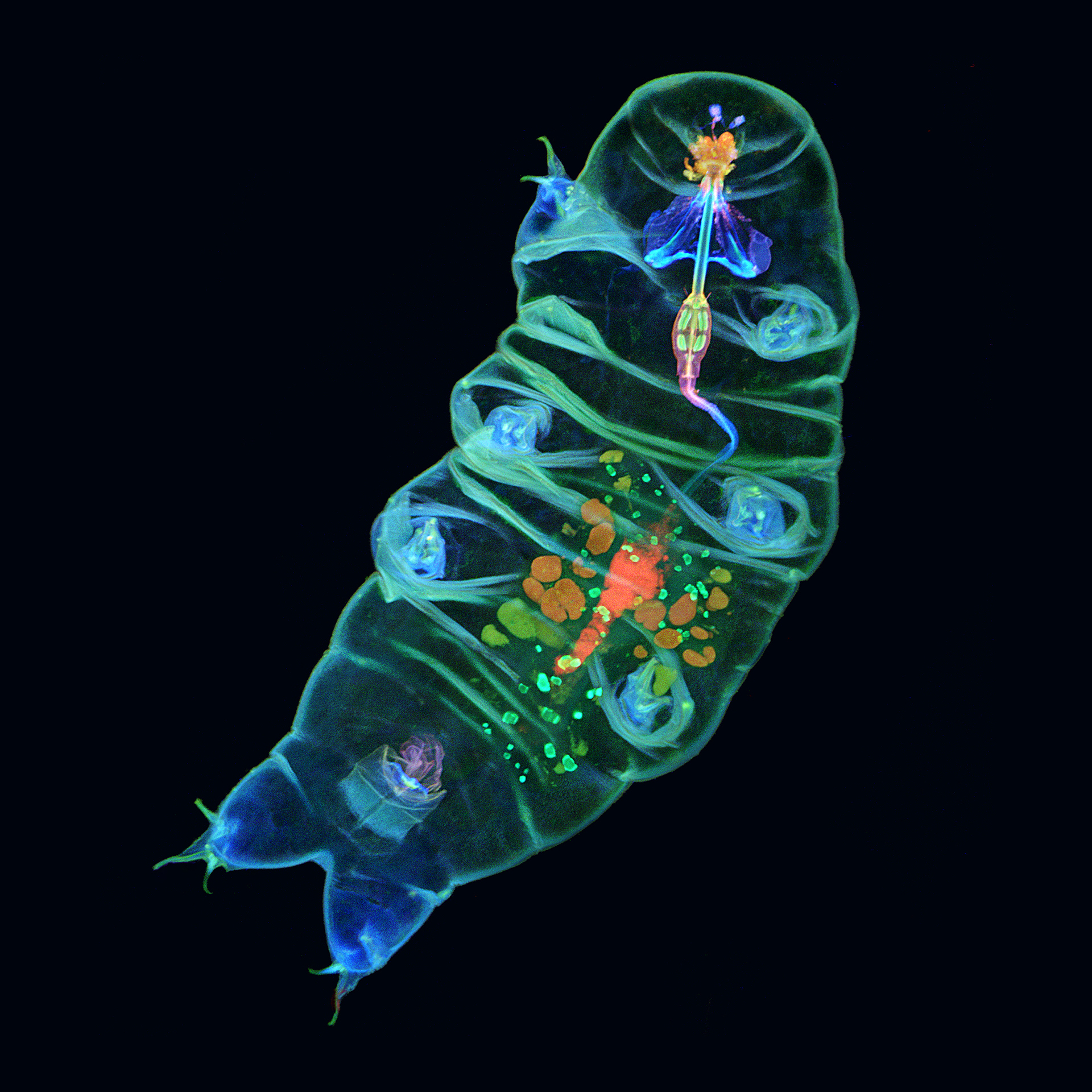 | Europe, the Middle East and Africa (EMEA) | Asia-Pacific |
Das Siegerbild wurde von Tagide deCarvalho (USA) aufgenommen. Das wunderschöne Fluoreszenzbild zeigt das Innere eines Bärtierchens mit farbenfrohen Details. | Das Bild von Alan Prescott (UK) gewann den EMEA-Regionalpreis. Sein Bild trägt den Titel „The mouse’s whiskers“ (Schnurrhaare einer Maus) und ist ein Gefrierschnitt einer Maus unter Verwendung mehrerer Fluoreszenzmarker. | Der Preis der Region Asia-Pacific ging an Howard Vidin (Australien). Dieses bemerkenswerte Bild zeigt die Autofluoreszenz eines Mausembryos mit 950 zusammengesetzten Kacheln. |
Ehrenvolle Erwähnungen
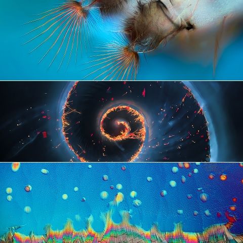
 Dies sind Eizellen im Eierstock einer galleninduzierenden Wespe, Anselmella miltoni Girault, aufgenommen mit einem konfokalen Mikroskop. |  Blütenstand von Arabidopsis thaliana mit jungen, sich entwickelnden Blütenknospen, die einen Fluoreszenzreporter exprimieren. |  Ein Präparat der Aminosäuren L-Glutamin und Beta-Alanin, das aus einer Ethanollösung kristallisiert und bei 50X mit Polarisationsfiltern aufgenommen wurde. |
 Rückenmark einer Maus mit einer GFP-Expression und geklärt mit der CLARITY Methode. |  Bild mit 3D-Tiefe: farbcodierte Rekonstruktion konfokaler Bilder von Tubulin (Mikrotubuli) in mitotischen COS3-Affenfibroblasten. |  Es wurde ein Teil der faltbaren Flügel eines Insekts aufgenommen mit dem Titel „A road in the sky“, da die Adern wie Straßen und die Stacheln der Flügelmembran wie Sterne aussehen. |
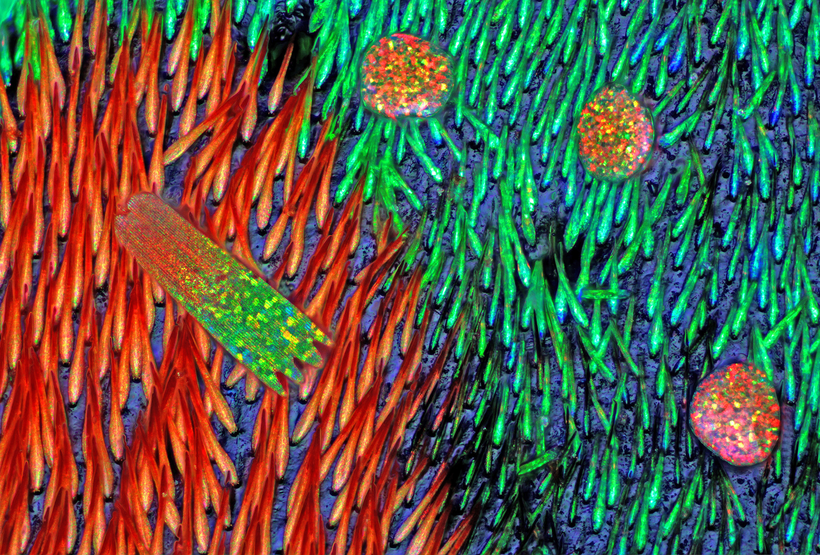 Verschiedene photonische Kristalle in Insekten auf dem Elytron des Bockkäfers Sternotomis pulchra. | 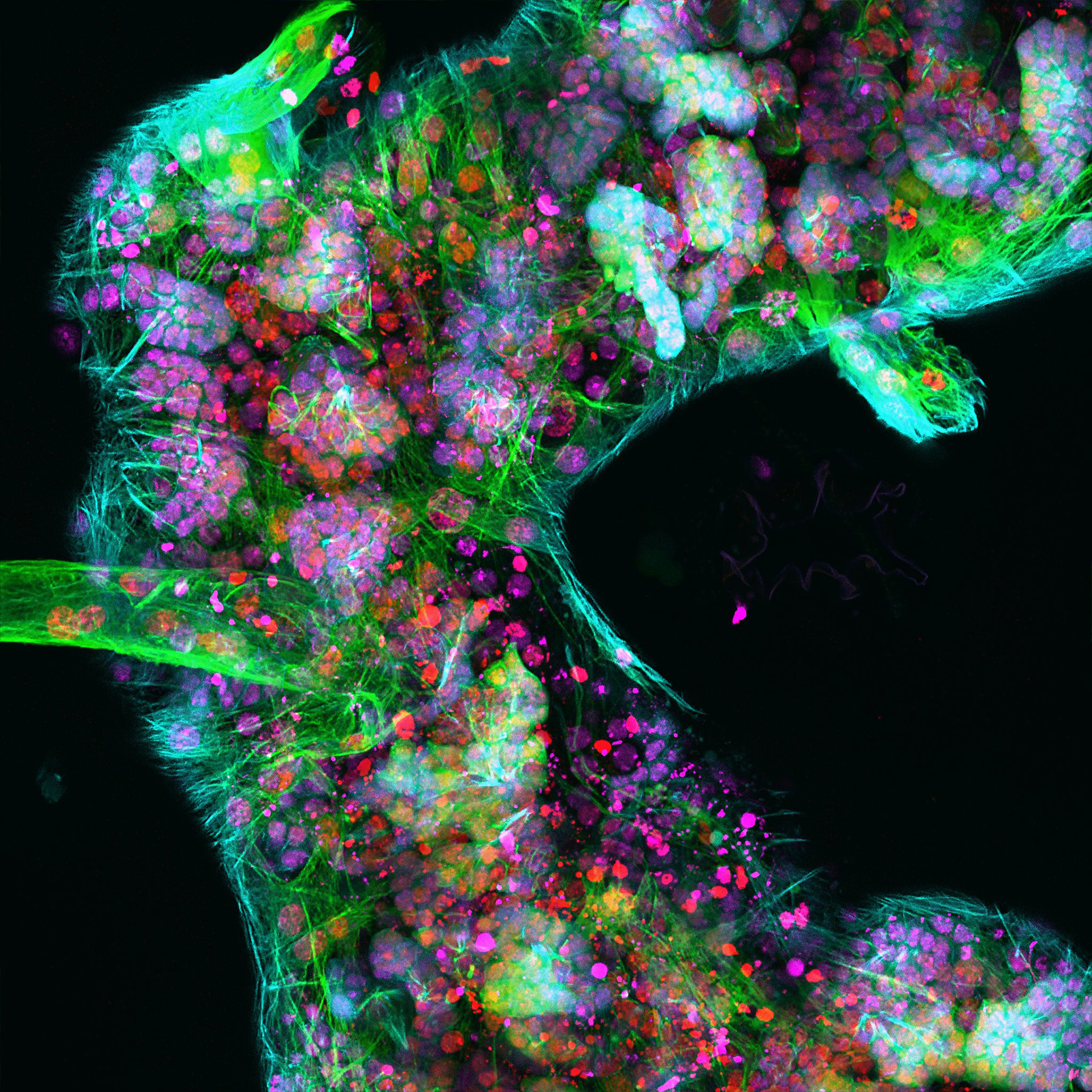 Zellkerne (rot) und Mikrotubuli (grün) markieren die Neuronen des Gehirns. | 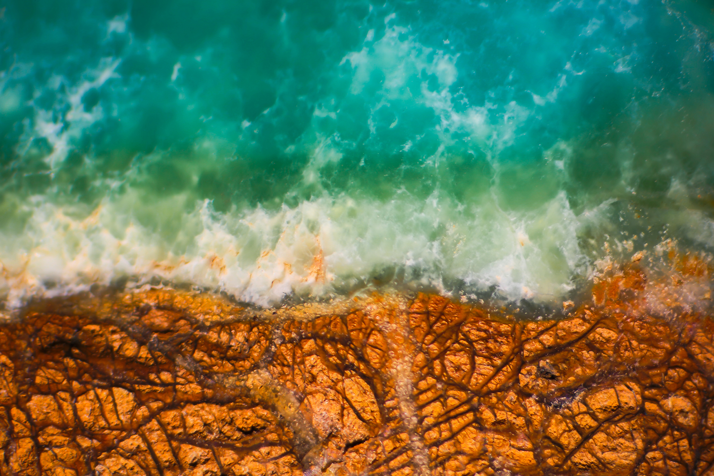 Dieses Bild zeigt das grüne Edelsteinmaterial Prase-Opal, das bei Vergrößerung durch ein Mikroskop bemerkenswert an eine Luftaufnahme einer Küstenlinie erinnert. |
Hintergrundbilder für Desktop als Download
Laden Sie das Bildpaket des Image of the Year Award jetzt kostenlos als Desktophintergrund herunter und verschönern Sie Ihren Bildschirm!
Hintergrundbildpaket für Desktop als Download (ZIP, 33.8 MB)
Hintergrundbildpaket für Mobiltelefon als Download (ZIP, 23.7 MB)
Sorry, this page is not
available in your country.
Sorry, this page is not
available in your country.