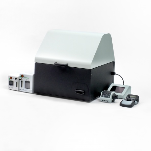
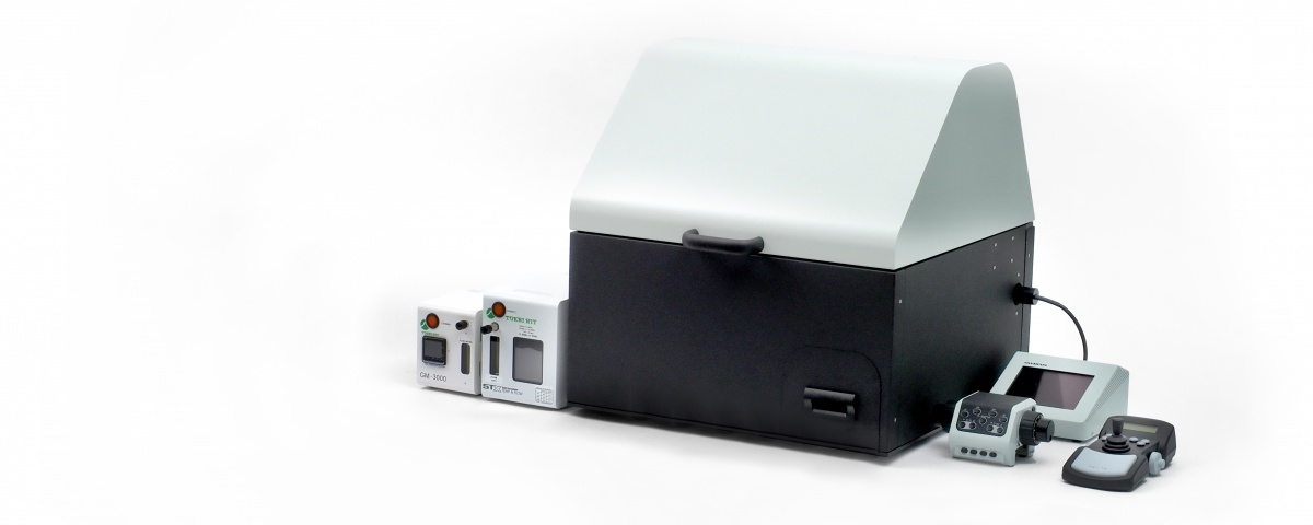
Bioluminescence imaging at the single-cell level enables you to validate a preliminary evaluation of luminescence assays using a plate reader, and clarifies the causes of unexpected results during experiments. Our IXplore™ Live for Luminescence microscope system* is optimized for both single-cell and whole tissue bioluminescence imaging. The microscope comes with the necessary components to begin your bioluminescence experiments immediately in a stable, controlled environment.
*This system was developed in collaboration with Professir Takeharu Nagai, Sanken, Osaka University, Tokai Hit Co. Ltd. and Evident. The project was funded by the Japan Science and Technology Agency (JST).
Observe Multiple Samples in a Microplate
Acquire images reliably across a variety of microplate designs at a resolution that can identify cells. cellSens™ imaging software and a motorized stage enable the system to memorize the XY position of each well, making it easy to capture images of all wells in a 96-well plate.
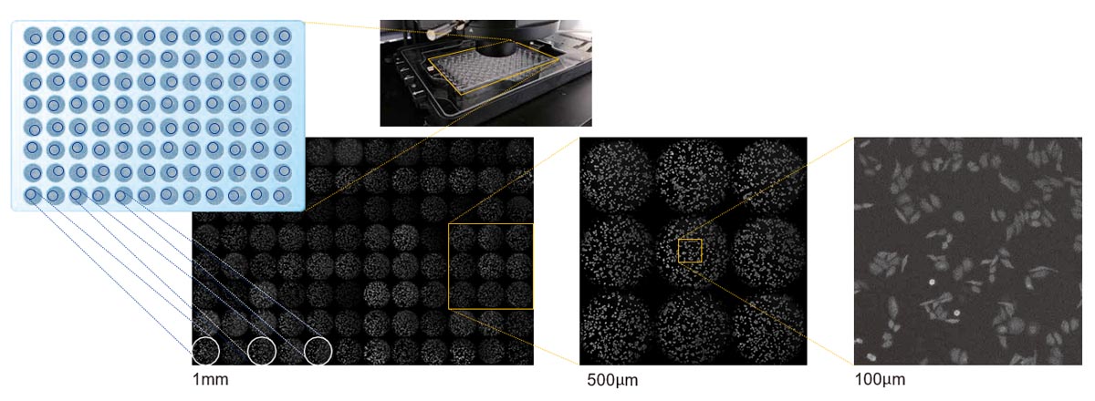
Sample: HeLa cells expressing yellow-enhanced Nano-Lantern. Data courtesy of Takeharu Nagai, Mitsuru Hattori, Department of Biomolecular Science and Engineering, Sanken, Osaka University.
Stable and Long-Term Time-Lapse Imaging
An on-stage incubation system provides the proper environment for long-term timelapse experiments of live cells and tissues. The system controls the temperature, CO₂, and other environmental conditions to maintain the health of your samples during imaging.
Bioluminescence imaging does not require excitation light like fluorescence since the source of light is a chemical reaction. Bioluminescence minimizes phototoxicity and eliminates autofluorescence signal contribution, making it ideal for stable, long-term observation of living cells.
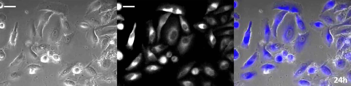
Long-term observation using an on-stage incubator (24 hours: phase contrast / luminescence / merge). Sample: HeLa cells expressing yellow-enhanced Nano-Lantern (35 mm glass bottom dish).
Data courtesy of Takeharu Nagai, Mitsuru Hattori, Department of Biomolecular Science and Engineering, Sanken, Osaka University.
Combine Fluorescence, Bioluminescence, and Transmitted Light Imaging
Combine observation methods and modalities to see changes in the entire cell and at the gene level at the same time. For instance, observe Ascl1 gene expression and the cell cycle in the self-replication process of neural stem cells. By using a fluorescent ubiquitination-based cell cycle indicator (Fucci), the system enables you to visualize the fluctuations of Ascl1 gene expression in each cell cycle stage.
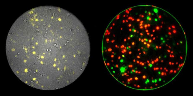
Three-channel single-cell resolution imaging with fluorescence (right, red/green), bioluminescence (left, yellow), and phase contrast (left, grayscale). Data courtesy of Itaru Imayoshi, Research Center for Dynamic Living Systems, Graduate School of Biostudies, Kyoto University; Akihiro Isomura, Ryoichiro Kageyama, Institute for Virus Research, Kyoto University.
Reference: Science. 2013 Dec 6;342(6163):1203-8. doi: 10.1126/science.1242366.
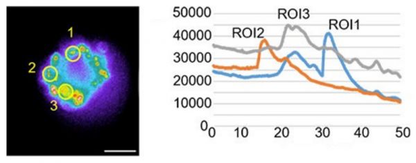
Drug Response Imaging for Efficacy Assessment
The activity of G protein-coupled receptors (GPCRs)—a commonly studied drug target—can be measured using the calcium concentration in a spheroid. High-content screening and analysis using a microplate enables you to analyze the different conditions, such as concentrations and environment conditions, as well as the library of the drug candidates.
A stable incubation system lets you observe cell morphological differentiations and the response to the drugs over longer periods of time. Bioluminescence has a better signal-to-noise (SNR) ratio compared to fluorescence, so you can detect even minute responses.
Explore the various bioluminescence imaging applications possible with the system and reach out to our experts for more information. |
|---|
Sorry, this page is not
available in your country.
Sorry, this page is not
available in your country.