Keep Your Samples Moving
Easy to operate, the automated motorized stage tracking software
for FLUOVIEW systems detects the target object in relation to the XY stage’s
centroid position during time-lapse image acquisition and moves the stage
to keep the target object near the center of the field of view.
Automated Motorized Stage TrackingKeeping moving samples, like C. elegans, in a microscope’s field of view during time-lapse imaging is challenging. Our automated motorized stage tracking software for FLUOVIEW laser scanning microscopes helps solve this challenge. Easy to operate, the software detects the target object in relation to the XY stage’s centroid position during time-lapse image acquisition and moves the stage to keep the target object near the center of the field of view. With two detection algorithms for fluorescence and transmission images, the software is designed for XYT/XYZT* time-lapse imaging. *Only the horizontal direction (XY coordinates) is automatically adjusted in this software | |
| Image data are courtesy of the following institution:
Sample condition: C. elegans labeled green (muscle cells, myo-3p::GCaMP3) and red (coelomocyte, unc-119p::dsRed)
|
| Trajectory InformationAt the end of an acquisition, the software outputs trajectory information (log data of the acquisition time and stage XY coordinates) as a CSV file along with the image data. This data can be used as part of a behavioral analysis by graphing it using Microsoft Excel or a similar program. For challenging samples, you can enable the software’s Manual mode. In this mode, you can click on the target object in the live image to track it. Like Auto mode, the image data and trajectory information can easily be exported. |
|---|
| "We were in dire need of tracking software that could acquire multicolor fluorescent images of neurons while automatically tracking fast-moving C. elegans.
Asuka Takeishi, Ph.D. |
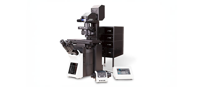
Supported Microscope Systems
Confocal Laser Scanning Microscope FV4000
*As of October 2023. |
Sorry, this page is not
available in your country.
Sorry, this page is not
available in your country.